37 blind spot ray diagram
Main Problem With Spot Diagrams • The main problem is that spots in the spot diagram don't convey intensity – A ray intersection point in the diagram does not tell the intensity at that point 0.000,0.000 DG 0.00, 0.00 0.000,10.00 DG 0.00, 0.71 0.000,14.00 DG 0.00, 1.00 FIELD POSITION DEFOCUSING 0.00000 Double Gauss - U.S. Patent 2,532,751 ... A useful technique for discovering blind spots is the Johari Window. Created in 1955 by Joseph Luft and Harry Ingham, the model is used to help individuals better understand themselves and how they are perceived by others. The window consists of four segments (or panes) of human interaction: open, hidden, blind and unknown.
LANL Engineering Standards Manual PD342 Chapter 17 Pressure Safety Section D20-B31.3-G, ASME B31.3 Process Piping Guide Rev. 2, 3/10/09 4 The Owner and Designer are responsible for compliance with the personnel and process qualification requirements of the codes and standards. In particular, the application of ASME B31.3 requires compliance with the Inspector qualification

Blind spot ray diagram
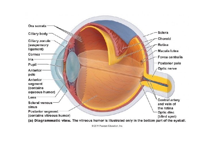
The Blind Spot. There is something kind of funky about the setup here. The photoreceptors are at the back of the retina, the ganglion cells are at the front, and the ganglion cell axons make up the optic nerve that goes out through a hole at the back. There can't be any photoreceptors here where the hole is. The Blind Spot One of the most dramatic experiments to perform is the demonstration of the blind spot. The blind spot is the area on the retina without receptors that respond to light. Therefore an image that falls on this region will NOT be seen. It is in this region that the optic nerve exits the eye on its way to the brain. Blind Spot, by Linda Shore ... Your blind spot is a hole in your retina where the optic nerve enters the back of ... Figure 5 also shows a ray diagram that.4 pages
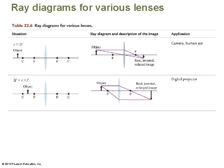
Blind spot ray diagram. 0:11Equipment blind spots are dynamic! By overlaying a 3D point cloud from a laser scan of a construction forklift ...20 Sep 2016 · Uploaded by Jochen Teizer Ray Diagram for Object Located at the Focal Point Thus far we have seen via ray diagrams that a real image is produced when an object is located more than one focal length from a converging lens; and a virtual image is formed when an object is located less than one focal length from a converging lens (i.e., in front of F ). 2 Mar 2020 — Answer · 1. Draw the image of the object. · 2. Pick one extreme on the image of the object and draw the reflected ray that will travel to the eye ...2 answers · Top answer: Answer:[tex]\huge{\fbox{\fbox{\bigstar{\mathfrak{ ed{answer}}}}}}[/tex]Here's ... LANL Engineering Standards Manual STD-342-100 Chapter 17-Pressure Safety Section REF References Rev. 0, 09/17/2014 REF-3 ASME B31.3 Process Piping Guide 1 of 171 . ASME B31.3 Process Piping Guide
by J Sanny · Cited by 1 — mine the diameter of their “blind spot.” The level of the calculation is ... asterisk diagram and the meterstick. ... the light ray from. A blind spot, scotoma, is an obscuration of the visual field. A particular blind spot known as the physiological blind spot, "blind point", or punctum ... 24 Aug 2011 — Diagram: blind spots while driving ... This is an experiment with what I can learn from interactive diagrams. There are several diagrams showing ... Science activity demonstrating the blind spot phenomenon ... spot on the light-sensitive lining, or retina, of your eye (click to enlarge diagram below).
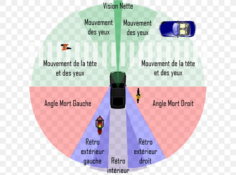
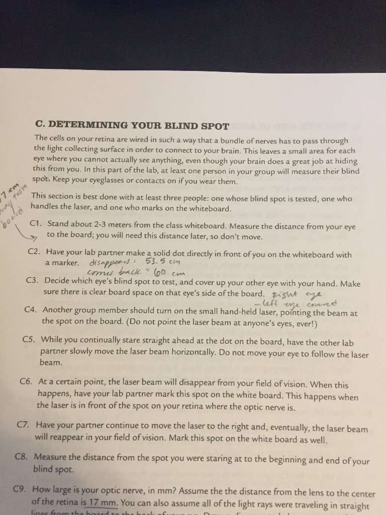
Every human eye has something called a blind spot. This natural blind spot is the place in the retina — the light-sensitive inner lining at the back of your eye — that doesn’t have any cells that respond to light. The blind spot sits in the part of your retina where the optic nerve exits the eye.. Why do you have blind spots? Blind spots are a normal part of your vision. Transfers – Shuttle Buggies. Roadtec 2500B. Show More. Disclaimer: The blind area diagrams are reproduced as received from the contractor, Caterpillar Inc., under NIOSH contract number 200-2002-00563. The opinions, findings, and conclusions expressed herein are not necessarily those of the National Institute for Occupational Safety and Health ... Please help! I am not good a drawing ray diagrams, and I would like to double check what I did to make sure its right. Thanks! a) Draw a ray diagram of the experiment conducted in part C (determining your blinds spot) only include rays that travel straight from the laser on the board to your eye. b) Draw a ray diagram of the lens system in part D. The ray diagram in Figure 2 shows image formation by the cornea and lens of the ... (a) Light rays from the same point on a distant object must be nearly ...

Blind Spot
Blind Spot, by Linda Shore ... Your blind spot is a hole in your retina where the optic nerve enters the back of ... Figure 5 also shows a ray diagram that.4 pages

Cadillac Camera Mirror Sees Through Your Car Roadshow
The Blind Spot One of the most dramatic experiments to perform is the demonstration of the blind spot. The blind spot is the area on the retina without receptors that respond to light. Therefore an image that falls on this region will NOT be seen. It is in this region that the optic nerve exits the eye on its way to the brain.

Alexis Work
The Blind Spot. There is something kind of funky about the setup here. The photoreceptors are at the back of the retina, the ganglion cells are at the front, and the ganglion cell axons make up the optic nerve that goes out through a hole at the back. There can't be any photoreceptors here where the hole is.

16 3 Lenses Texas Gateway
2
Pdf Using Population Models To Validate Platzer S Methodology For Overcoming Vehicle Side Mirror Blind Spots Semantic Scholar
1

The Human Eye And The Colourful World Lakhmir Singh Manjit Kaur Solutions Pg No 271 Class 10 Physics

Diagram Showing Cross Section Of Human Eye Stock Vector Illustration Of Child Sight 158742341

Anterior Blind Area Of The Avian Visual Field A Side View Of A Download Scientific Diagram
With The Help Of A Neat And Labelled Diagram Describe The Anatomy Of Human Eye Explain The Mechanism Of Vision Sarthaks Econnect Largest Online Education Community
The Car Guide Vehicle Blind Spot Visual Field Visual Perception Png 650x609px Car Driving Eye Joint

Blind Spot Visualization A Blind Spot Visualization Potentially Download Scientific Diagram
2

Stereo Panoramic Vision For Monitoring Vehicle Blind Spots Ppt Download
Cyberphysics Depth Of Object Field And Depth Of Image Focus

Sangat Ringan Titanium Murni Bingkai Bundar Kacamata Minus Wanita Pelindung Mata Anti Blu Ray Anti Kelelahan Modis Makeup Tidak Berderajat Blind Spot Mirror Mata Lazada Indonesia

Blind Spot Vision Wikipedia
2
Mirrors And Lenses How Do Eyeglasses Correct Your

Eye And Ear
2

Ocular Anatomy And Optics Sharks Rays And Chimaeras
The Human Eye At School You Are Taught

Sciencoknowledge Spherical Mirrors

Automating The Blind Spot Measurement Of Construction Equipment Sciencedirect

Model Question Paper 3
Volvo S60 Volvo Cars Global Media Newsroom

Gcse Biology Eye Diagram Diagram Quizlet

12 6 How Do We See Light Visible Light Siyavula
Mirrors And Lenses How Do Eyeglasses Correct Your

Human Eye In Cross Section

Ex4su01
2

Vehicle Blind Spot Wikipedia
Please Help I Am Not Good A Drawing Ray Diagrams Chegg Com

Reflection Ray Diagram For Mirror Image Applications Of Reflection Ppt Download
0 Response to "37 blind spot ray diagram"
Post a Comment