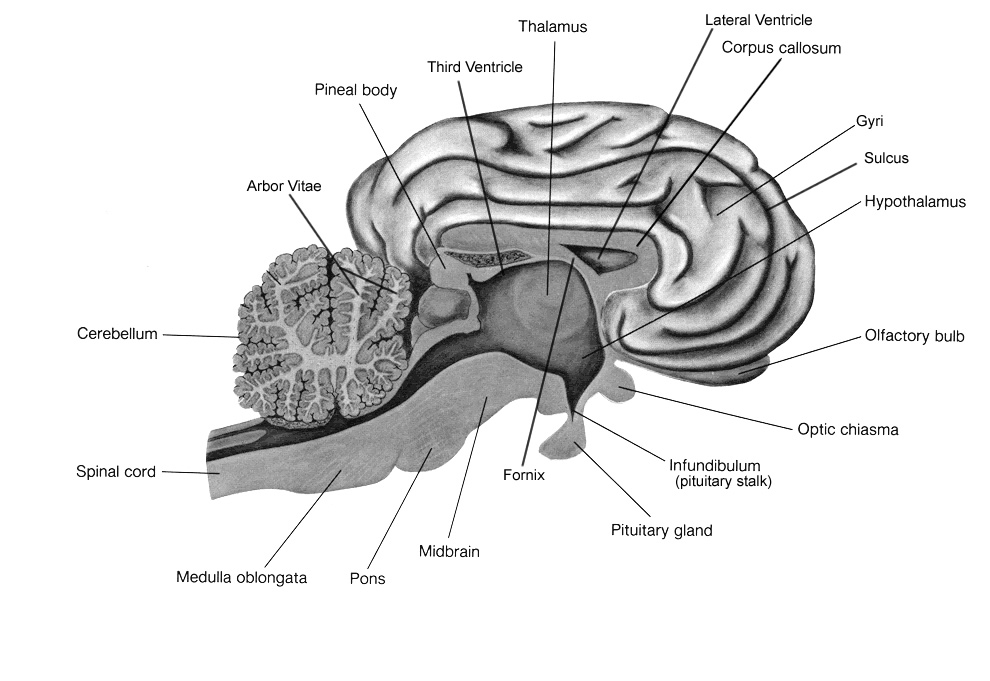
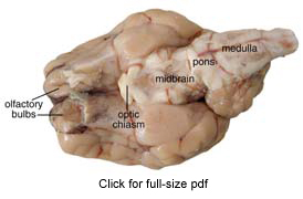
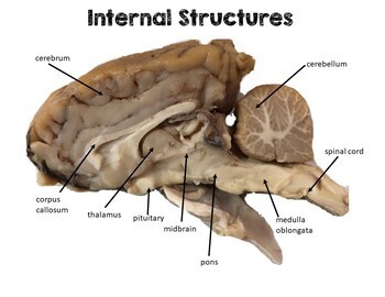
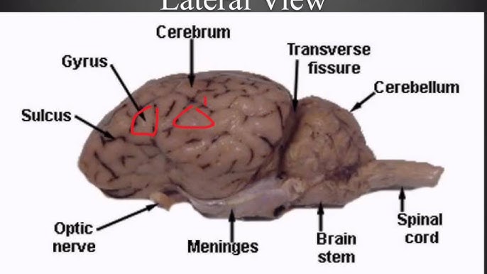
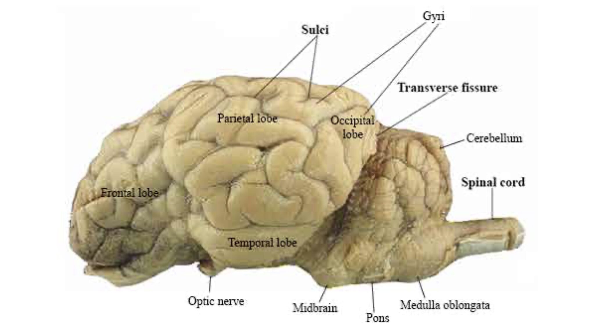
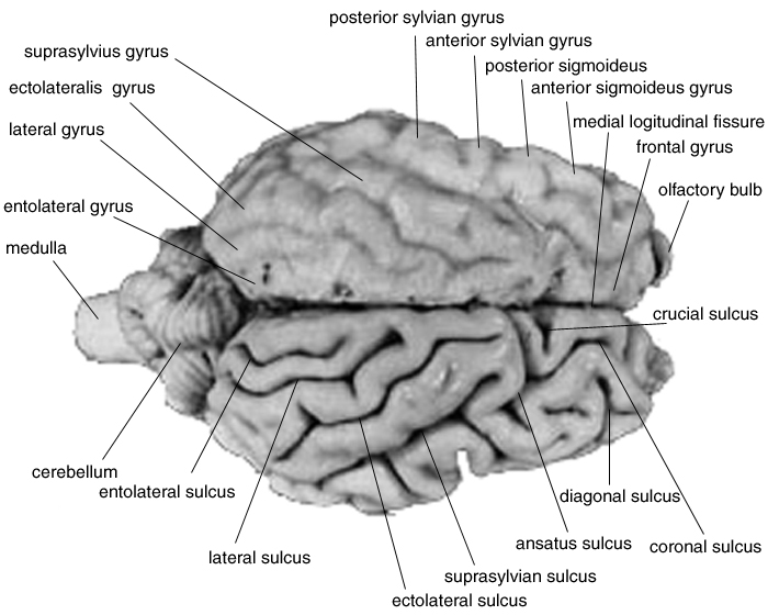
39 sheep brain diagram labeled
Sheep Brain Quiz - PurposeGames.com About this Quiz. This is an online quiz called Sheep Brain. This quiz has tags. Click on the tags below to find other quizzes on the same subject. Anatomy. sheep brain. Solved Art-labeling Activity: Midsagittal Section of the ... Anatomy and Physiology. Anatomy and Physiology questions and answers. Art-labeling Activity: Midsagittal Section of the Sheep Brain (Diagram, 2 of 2) Reset Help Fomix Infundibulum Olfactory bulb Optic chiasm Mosencephalon Pituitary gland Marillary body Medulla oblongata Pons Spinal cord Corpus callosum Art-labeling Activity: Midsagittal Section ...
Sheep Brain Label | Dissection, Human brain diagram, Brain ... Sheep Brain Label A drawing of the brain with the parts unlabeled. Students can practice naming the parts of the brain, then check their answers with the provided key. Biologycorner 17k followers More information unlabeled brain Find this Pin and more on A&P by Dijana Kovacevic. Brain Gym For Kids Human Brain Diagram Brain Anatomy And Function
Sheep brain diagram labeled
Label Sheep Brain Diagram | Quizlet Start studying Label Sheep Brain. Learn vocabulary, terms, and more with flashcards, games, and other study tools. Sheep brain labeling Quiz - PurposeGames.com The world - Fifteen islands 15p Image Quiz. Continue to count 15p Multiple-Choice. Midwest States and Capitals 12p Matching Game. Geometric Shapes 14p Image Quiz. Objects in ABBA songs - easy 2 15p Image Quiz. PG Lightning Game: Speed counting 20p Shape Quiz. States Without the Letter 'A' 14p Type-the-Answer. The Western States 11p Image Quiz. labeled brain - Pinterest Images taken from the dissection of the sheep's brain: cerebrum, cerebellum, corpus callosum, lobes, sulci, gyrus, fornix, pituitary.
Sheep brain diagram labeled. Sheep Brain Dissection Guide with Pictures | Nervous ... Dissection guide with instructions for dissecting a sheep brain. Checkboxes are used to keep track of progress and each structure that can be found is described with its location in relation to other structures. An image of the brain is included to help students find the structures. S La Luna Anatomy Neuron Diagram Science Biology Sheep Brain Dissection labeled 2 Diagram - Quizlet Sheep Brain Dissection labeled 2 STUDY Flashcards Write Test PLAY Match Created by AllieKlinger Terms in this set (8) Superior Colliculus Pineal Gland Cerebrum Thalamus Hypothalamus Cerebral Peduncle Pons Medulla Oblongata OTHER SETS BY THIS CREATOR Test Results4 Terms AllieKlinger Lecture 8 Part 225 Terms AllieKlinger Lecture 8 Part 114 Terms Sheep Brain Neuroanatomy Online Self-Test | KPU.ca ... Sheep Brain Neuroanatomy Online Self-Test. Use each diagram as a reference, and selected the correct answer for each lettered structure. You may find it useful to open the diagrams in a separate window to review while answering each question. Sheep Brain - San Diego Mesa College Sheep Brain. Click on a photo for a larger view of the model. Click on Label for the labeled model. Back to Dissected Specimen Page. Superior View. Corpora Quadrigemina. Label.
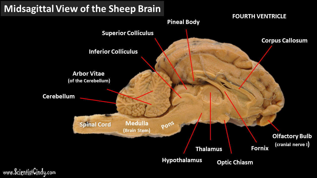
Sheep Brain Dissection Lab Use the picture below as a way to see how your sheep brain should look after you cut it in half. 10. In the image below, a probe indicates the location of the lateral ventricle. 11. Once the brain is cut this way, the colliculi can also be seen from the inside and the pineal gland is revealed only if you made a very careful incision. Diagram of Sheep Brain - Lateral view - Modesto Junior College Diagram of Sheep Brain - Lateral view - Modesto Junior College Sheep Brain Anatomy Diagram at Anatomy Image result for sheep brain labeled brain diagram. Source: labels-creative.com. Also, the sheep brain is oriented anterior to posterior (more horizontally), while the human brain is oriented superior to interior (more vertically.) materials. Almost all the basic task in the body is commanded by the brain. Fresh diagram of sheep brain. Sheep Brain Map: Midsaggital view This map shows the major structures of the sheep brain with an active cursor to help identify the structures Neuroanatomy Tutorial I: Basic Anatomy of the Brain Point to any region of this midsaggital sheep brain image (medial view) to highlight that structure. Click the left mouse button to identify the structure you are pointing to.
PDF Neuroanatomy: Dissection of the Sheep Brain The basic neuroanatomy of the mammalian brain is similar for all species. Instead of using a rodent brain (too small) or a human brain (no volunteer donors), we will study neuroanatomy by examining the brain of the sheep. We will be looking as several structures within the central nervous system (CNS), which is composed of the brain and spinal ... Sheep Brain - Veterinary Anatomy Website Home Page Dissected Sheep Brain — Dorsal View Dorsal view of sheep brain with the cerebellum and caudal cerebrum removed. The rostral colliculus(large arrow label) and the caudal colliculus(small arrow label) together form the tectumof the midbrain. PDF DISSECTION OF THE SHEEP'S BRAIN - Hanover College DISSECTION OF THE SHEEP'S BRAIN Introduction The purpose of the sheep brain dissection is to familiarize you with the three-dimensional structure of the brain and teach you one of the great methods of studying the brain: looking at its structure. One of the great truths of studying biology is the saying that "anatomy precedes physiology". Sheep Brain Dissection - Carolina.com Carolina's Perfect Solution® sheep brain dissection introduces students to the anatomy of a mammalian brain, both external and internal, and encourages students to construct an explanation of the central nervous system. Below is a brief survey of the internal and external anatomy of the sheep brain.
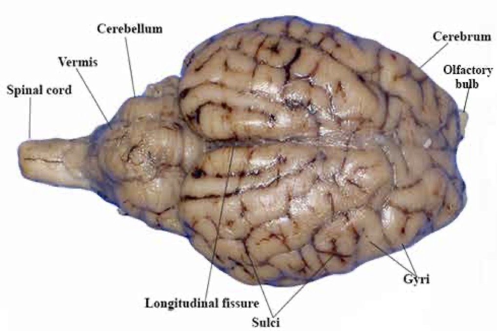
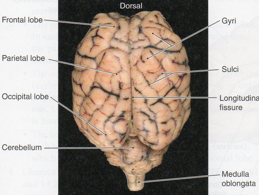
PDF Sheep Neuroanatomy Lab- Labeling Worksheet Psychology 2315 ... Sheep Neuroanatomy Lab- Labeling Worksheet Psychology 2315- Brain and Behaviour Kwantlen Polytechnic University Figure 1: Dorsal view Cerebellum, Frontal lobe, Occipital lobe, Parietal lobe, and Temporal lobe. Temporal Parietal Lobe Frontal Lobe Cerebellum Occipital Lobe
PDF Sheep Brain Dissection Lab - Home Science Tools Diagram Worksheets Print out the diagrams on the following pages and fill in the labels to test your knowledge of sheep brain anatomy. • External anatomy: label the top view (.pdf) • External anatomy: label the bottom view (.pdf) • Internal anatomy: label the right side (.pdf) See our other free dissection guides with photos and printable ...
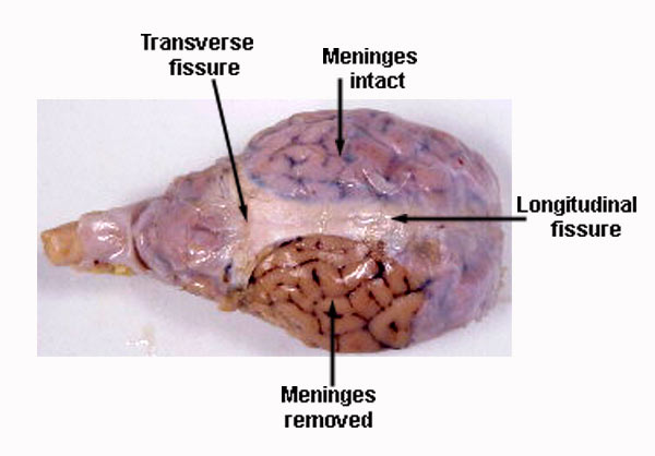
Sheep Brain Dissection with Labeled Images 1. The sheep brain is enclosed in a tough outer covering called the dura mater. You can still see some structures on the brain before you remove the dura mater. Take special note of the pituitary gland and the optic chiasma. These two structures will likely be pulled off when you remove the dura mater. Brain with Dura Mater Intact
Sheep brain dissection | Human Anatomy and Physiology ... The tough outer covering of the sheep brain is the dura mater, the outermost meninges membrane covering the brain. Remove the dura mater to see most of the structures of the brain, but remove it carefully, so as to leave all the other structures beneath it intact. Removing the dura mater from the cerebellum at the back of the brain can be tricky.
Sheep Brain Dissection Project Guide | HST Learning Center Download: Sheep Brain Dissection Lab Observation: External Anatomy of Sheep Brain. 1. You'll need a preserved sheep brain for the dissection. Set the brain down so the flatter side, with the white spinal cord at one end, rests on the dissection pan. Notice that the brain has two halves or hemispheres.
Sheep Brain Anatomy and Function Flashcards - Cram.com Study Flashcards On Sheep Brain Anatomy and Function at Cram.com. Quickly memorize the terms, phrases and much more. Cram.com makes it easy to get the grade you want!
Sheep Brain Dissection labeled Diagram | Quizlet Only $2.99/month Sheep Brain Dissection labeled STUDY Learn Write Test PLAY Match Created by AllieKlinger Terms in this set (8) Corpus Collosum Lateral Ventricle Fornix Hypothalamus Cerebral Aqueduct Central Canal Inferior Collicuious Transverse Fissure THIS SET IS OFTEN IN FOLDERS WITH... Sheep Brain Dissection labeled 2 8 terms AllieKlinger
PDF Sheep Brain Midsagittal Section - Dr. Scott Croes' Website Sheep Brain –Midsagittal Section. Choroid Plexus of Lateral Ventricle Parietal Lobe Thalamus Cerebellum Frontal Lobe Pons Variollii Medulla Oblongata Optic Nerve Corpus Callosum Lateral Ventricle Lentiform Nucleus Hypothalamus Spinal Cord Hippocampus Superior Inferior Colliculus Colliculus Cerebral Peduncle Central Sulcus Arbor Vitae Occipital Fornix Pituitary Gland Parasagittal Section.
PDF Lab: Sheep Brain Dissection - Mrs. Moretz's Science Site to anatomy studies. See for yourself what the . cerebrum, cerebellum, spinal cord, gray matter, white matter, and other parts of the brain look like! Observation: External Anatomy . 1. You'll need a . preserved sheep brain. for the dissection. Set the brain down so the flatter side, with the white . spinal cord. at one end, rests on the ...
DOC Sheep Brain Anatomy Lab Manual - amherst.edu The cruciate fissure (labeled ansate sulcus in your photo atlas) is known in the human brain as the fissure of Rolando or central sulcus, and intersects the medial longitudinal fissure to mark off the anterior third of the cortex.
Sheep Brain Anatomy Quiz - ProProfs Quiz 1. What is the outer covering of the Sheep brain? A. Arachnoid Mater B. Pia Mater C. Medulla Oblongata D. Dura Mater 2. Which amongst the following is the largest structure in dorsal view? A. Pons B. Medulla Oblongata C. Cerebral cortex D. Hippocampus 3. What are the large folds that surround the cerebrum? A. Gyri B. Sulci C. Fissures D. Colliculus
Sheep Brain Label - The Biology Corner Sheep Brain Label. Publisher: Biologycorner.com; follow on Google+. This work is licensed under a Creative Commons Attribution-NonCommercial 3.0 Unported License . Brain Label Answer Key. Image adapted from a photograph of the sheep brain.
Sheep Brain Images - San Diego Mesa College Sheep Brain Unlabeled. Sheep Brain Leader-Lined. Sheep Brain Labeled. San Diego Mesa College 7250 Mesa College Drive San Diego, CA 92111-4998 Student Support San Diego Community College District San Diego City College San Diego Mesa College San Diego Miramar College San Diego Continuing Education.
11.7: Sheep Brain Dissection - Biology LibreTexts 8 Jul 2021 — Anatomy & Physiology one organ at a time...Sheep Brain Dissection may be downloaded from .
Week 7: Sheep Brains with Labeled Cuts | Northfield ... Week 7: Sheep Brains with Labeled Cuts. Sheep brains used in science classrooms such as ours are salvaged after sheep are slaughtered for their meat. Sheep brains are a good resource for teaching neuroscience because they have many similarities to human brains both in anatomy and function, and are more abundant. Side view.
labeled brain - Pinterest Images taken from the dissection of the sheep's brain: cerebrum, cerebellum, corpus callosum, lobes, sulci, gyrus, fornix, pituitary.
Sheep brain labeling Quiz - PurposeGames.com The world - Fifteen islands 15p Image Quiz. Continue to count 15p Multiple-Choice. Midwest States and Capitals 12p Matching Game. Geometric Shapes 14p Image Quiz. Objects in ABBA songs - easy 2 15p Image Quiz. PG Lightning Game: Speed counting 20p Shape Quiz. States Without the Letter 'A' 14p Type-the-Answer. The Western States 11p Image Quiz.
Label Sheep Brain Diagram | Quizlet Start studying Label Sheep Brain. Learn vocabulary, terms, and more with flashcards, games, and other study tools.









0 Response to "39 sheep brain diagram labeled"
Post a Comment