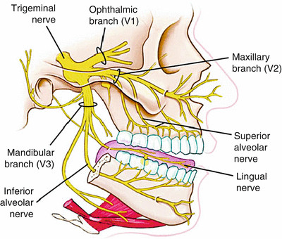
36 trigeminal nerve branches diagram
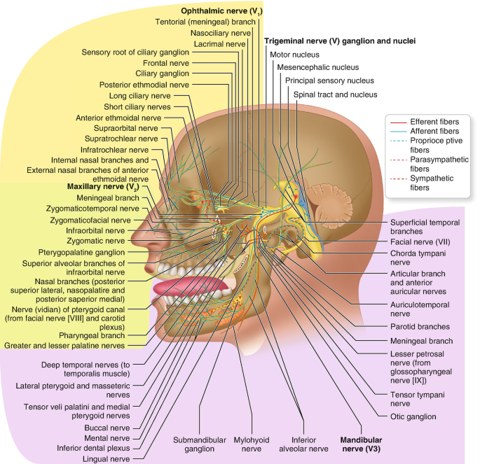
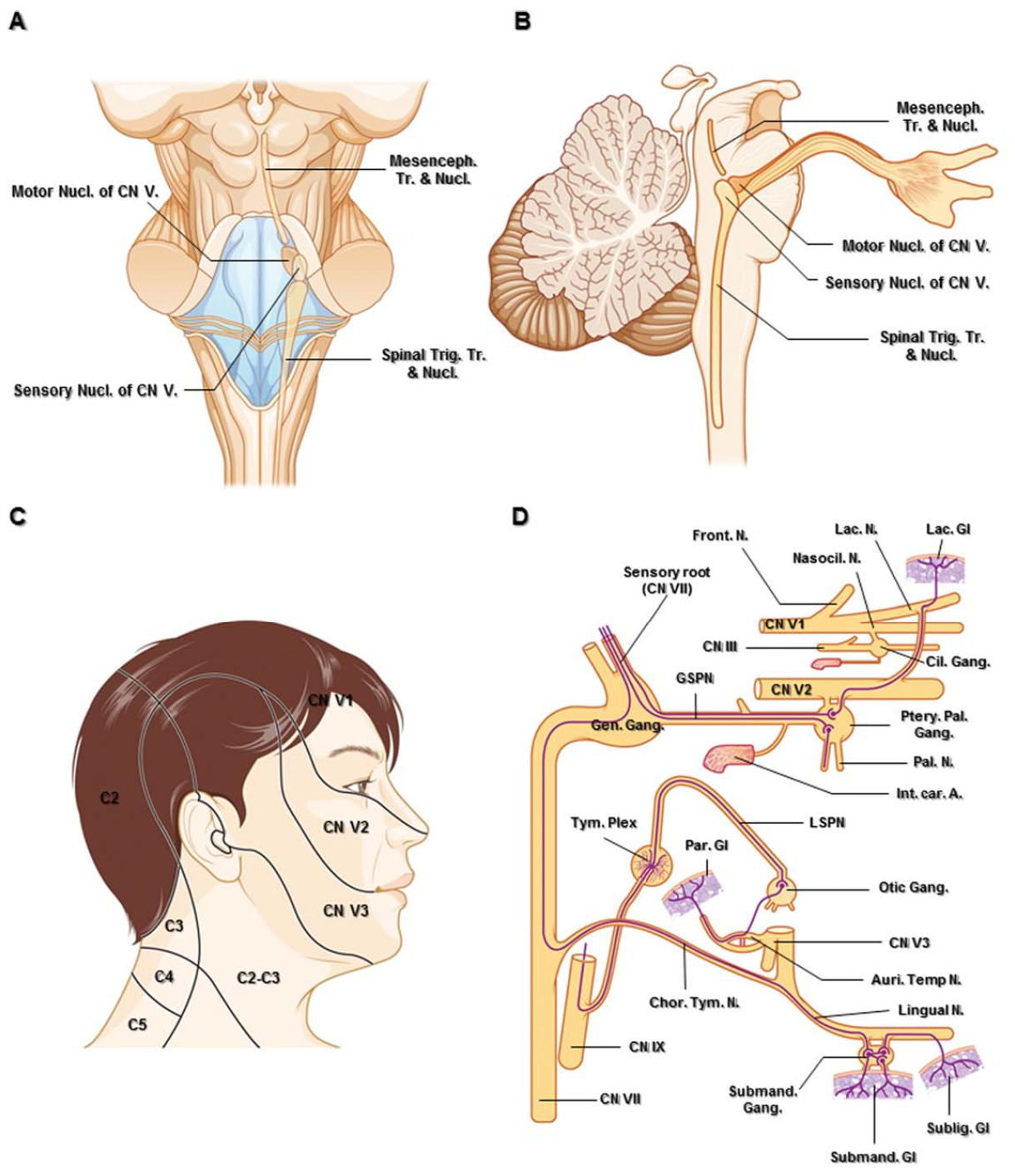
The Ophthalmic Division of the Trigeminal Nerve (CNV1 ... The cutaneous innervation to the face and scalp by the three branches of the trigeminal nerve have sharp borders and little overlap. The cutaneous innervation of CNV1 can be seen in the image below: Fig 4 - Cutaneous innervation to the head and neck. Autonomic Functions The ophthalmic nerve itself does not contain any autonomic fibres. Illustrations and diagrams of the 12 pairs of cranial ... The branches of the trigeminal nerve (V) are represented in three different diagrams The ophthalmic nerve (V1) in the orbital cavity with its main branches (frontal nerve, lacrimal nerve, anterior and posterior ethmoidal nerve, nasociliary nerve, branch communicating with the ciliary ganglion, supraorbital nerve, supratrochlear nerve, infra ...
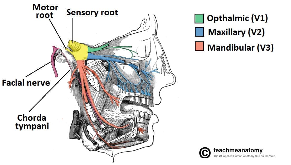
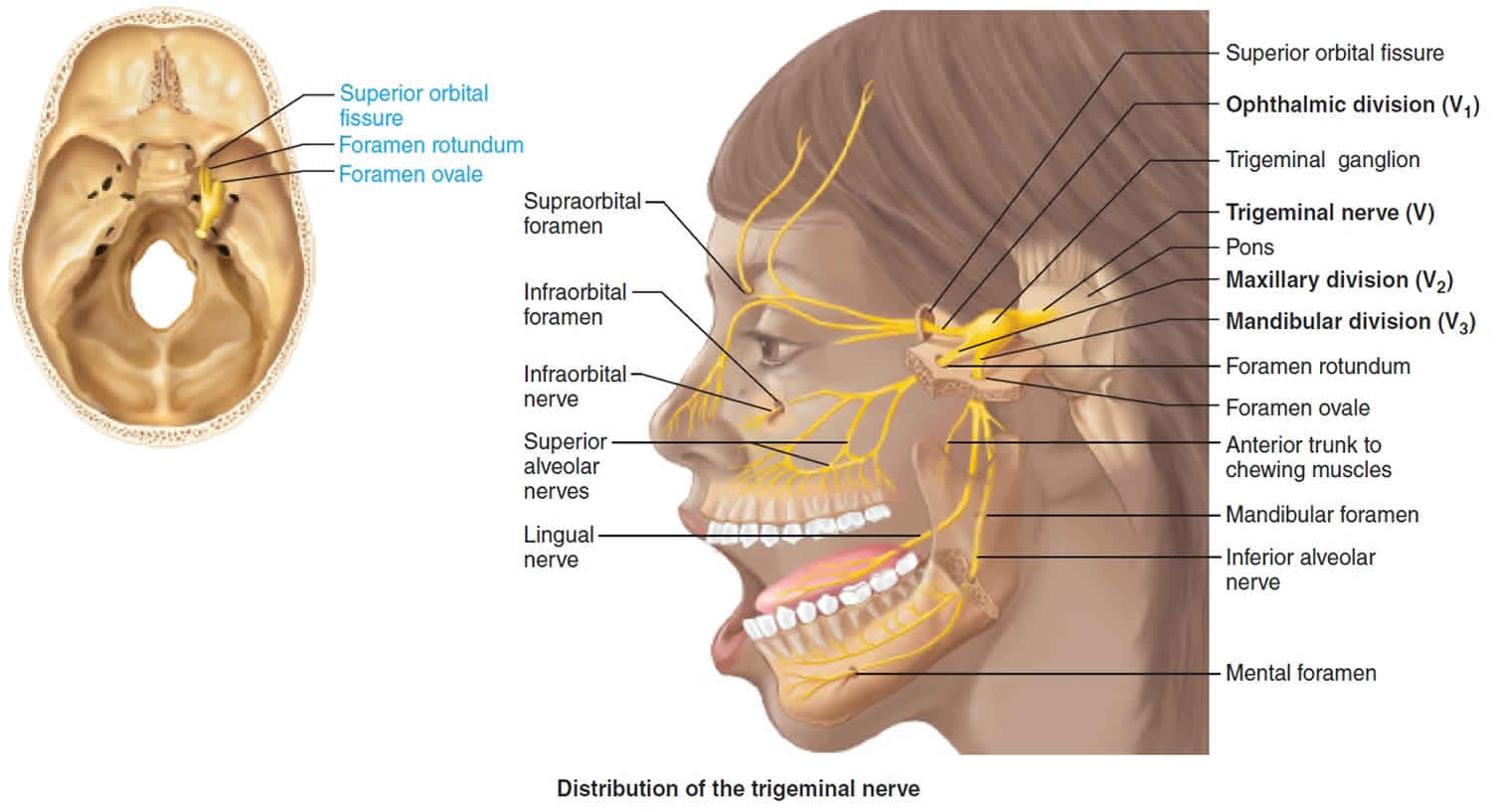
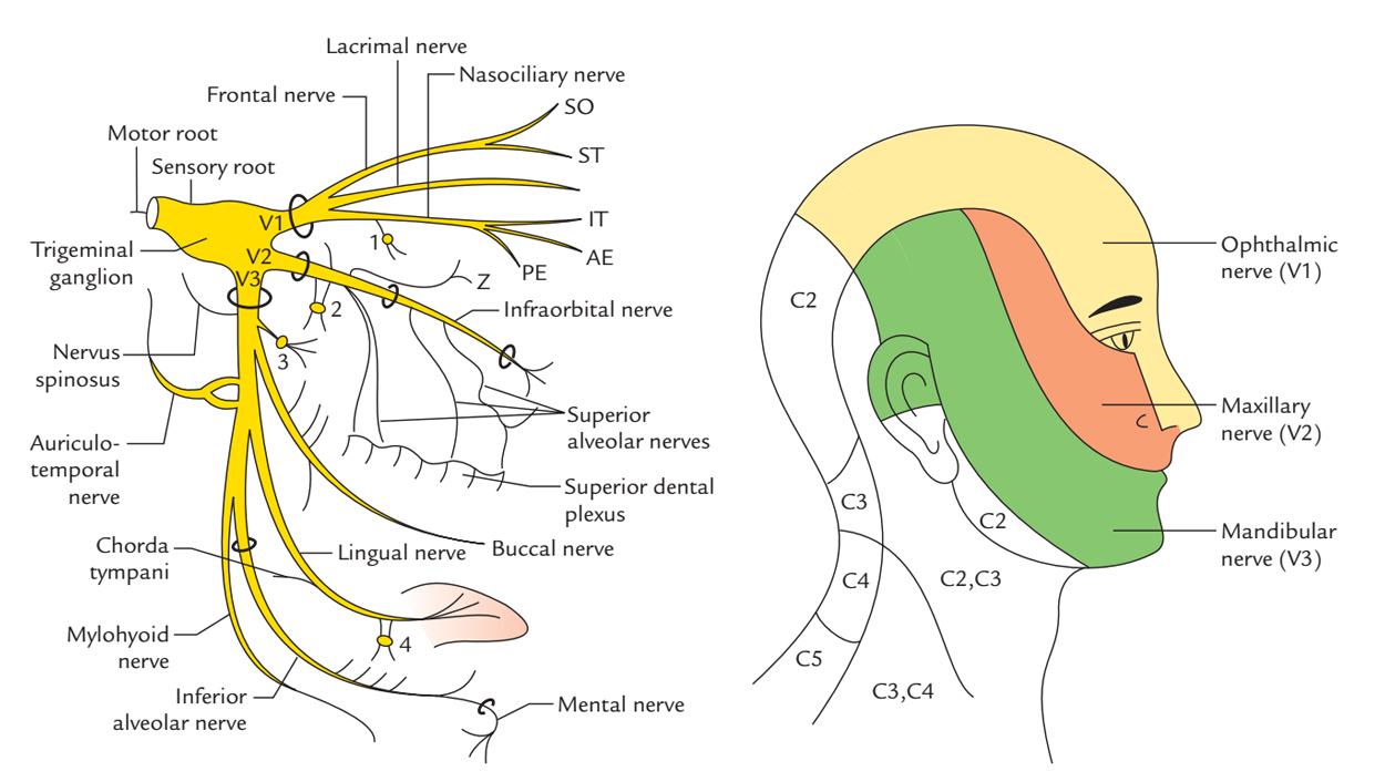
Cranial Nerve V: The Trigeminal Nerve - Clinical Methods ... The sensory portion of the trigeminal supplies touch-pain-temperature to the face. The nerve has three divisions: the ophthalmic, maxillary, and mandibular nerves (Figure 61.1). The innervation includes the cornea and conjunctiva of the eye; mucosa of the sinuses, nasal and oral cavities; and dura of the middle, anterior, and part of the posterior cranial fossae.
Trigeminal nerve branches diagram
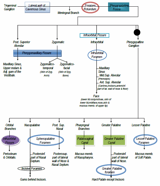
Trigeminal Nerve Anatomy Diagram shows trigeminal nerve (TGN), trigeminal ganglion, and peripheral divisions and their branches. From fora- men rotundum ossis sphenoidalis, maxil- lary nerve (thin underline) gains access to pterygopalatine fossa and continues in floor of orbit as infraorbital nerve. Inferior alveolar and lingual nerves ( thick underline ) The Trigeminal Nerve (CN V) | Cranial Nerves | Geeky Medics Cranial nerves and cranial foramina diagram. 1. Embryology. The name trigeminal is derived from the Latin "tri-" meaning three, and "-geminal" meaning a group attached to a common point. There are 'three' major branches of the trigeminal nerve, coming from four distinct nuclei in the brainstem. Trigeminal nerve (CN V): Anatomy, function and branches ... The branches of the ophthalmic division of the trigeminal nerve are summarized below. Learn the anatomy of ophthalmic nerve with our videos, quizzes, articles, and labeled diagrams: Ophthalmic nerve (CN V1) Explore study unit Maxillary division (CN V2)
Trigeminal nerve branches diagram. Trigeminal Nerve Anatomy: Gross Anatomy, Branches of the ... The semilunar (gasserian or trigeminal) ganglion is the great sensory ganglion of CN V. It contains the sensory cell bodies of the 3 branches of the trigeminal nerve (the ophthalmic, mandibular,... Trigeminal Neuralgia Fact Sheet | National Institute of ... The trigeminal nerve is one of 12 pairs of nerves that are attached to the brain. The nerve has three branches that conduct sensations from the upper, middle, and lower portions of the face, as well as the oral cavity, to the brain. The ophthalmic, or upper, branch supplies sensation to most of the scalp, forehead, and front of the head. Trigeminal Nerve Diagram - Trigeminal Neuralgia - MedHelp Thanks for the diagram. What I've learned over the last year and a half is that there are little branches from the TN nerve that go to each of the teeth -- that's why a lot of us experience 'toothache' like pain. And the diagram shows that it connects into the nose area -- that's where I had my first electric zap. PDF 5. THE TRIGEMINAL SYSTEM Somatic Sensation of the Face and ... 2. Three nerve roots give rise to: a. Ophthalmic nerve, (CN V-1) b. Maxillary nerve, (CN V-2) c. Mandibular nerve, (CN V-3) 3. Peripheral distribution of three branches. Back of head and the angle of the jaw are not supplied by the trigeminal (Areas around ear supplied by CNs
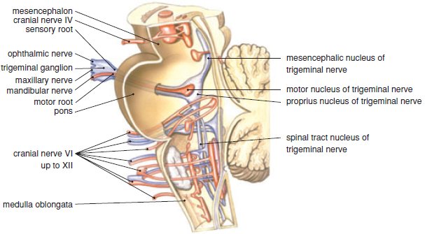
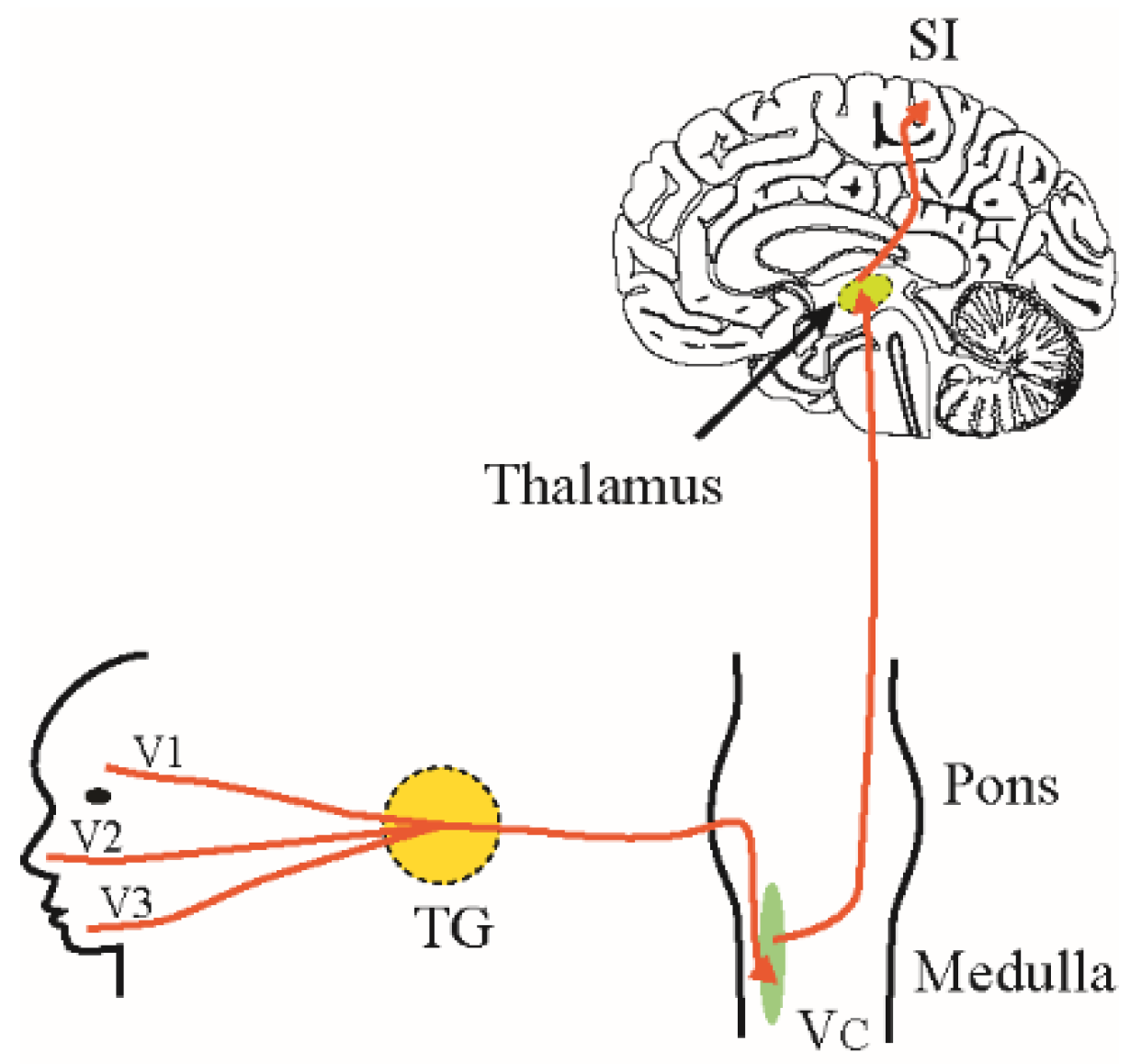
Neuroanatomy, Trigeminal Reflexes - StatPearls - NCBI ... The trigeminal nerve is the fifth cranial nerve (CN V) and the largest of the paired cranial nerves. It is a mixed nerve that partially innervates the craniofacial region along with the facial nerve. The fifth cranial nerve contains three terminal branches that innervate the skin of the face and neck, mucous membranes and paranasal sinuses of ... Trigeminal nerve (illustration) | Radiology Case ... Diagram Simplified diagrams of the main branches and sensory supply of the trigeminal nerve. Case Discussion Simplified illustrations of the trigeminal nerve, its main branches, and its sensory supply. See article for more information. References 11 article feature images from this case 1 public playlist includes this case Trigeminal Nerve Distribution Diagram | Quizlet Start studying Trigeminal Nerve Distribution. Learn vocabulary, terms, and more with flashcards, games, and other study tools. Trigeminal Nerve Anatomy : American Journal of ... —Sagittal diagram shows three peripheral divisions of trigeminal nerve entering convexity and root bundles leaving concavity of sickle-shaped trigeminal ganglion. Motor root (solid arrowhead) bypasses ganglion and reunites with mandibular nerve in foramen ovale basis cranii. Open arrowhead indicates descending spinal trigeminal tract.
Trigeminal Nerve: Function, Anatomy, and Diagram Explore the interactive 3-D diagram below to learn more about the trigeminal nerve. Testing The trigeminal nerve plays a role in many sensations that are felt in different parts of the face. As a... Pin by Beth Elizabeth Worley on Trigeminal Neuralgia ... Facial pain info, trigeminal neuralgia is an inflammation of the trigeminal nerve causing extreme pain and muscle spasms in the face. Causes, diagnosis and treatment. Kem Friddle. Health Information. Atypical Trigeminal Neuralgia. Peripheral Neuropathy. Rheumatoid Arthritis. Cranial Nerve 5. The Trigeminal Nerve (CN V) - Course - Divisions ... The trigeminal nerve, CN V, is the fifth paired cranial nerve. It is also the largest cranial nerve. In this article, we shall look at the anatomical course of the nerve, and the motor, sensory and parasympathetic functions of its terminal branches. The trigeminal nerve is associated with derivatives of the 1st pharyngeal arch. Oral Anatomy 2: Trigeminal Nerve Diagram | Quizlet What are the 3 branches of the trigeminal nerve? 1. Ophthalmic (sensory) 2. Maxillary (sensory) 3. Mandibular (mixed) Ophthalmic branch -afferent (sensory) -little dental significance; may have side effects if LA is given incorrectly -smallest branch The ophthalmic branch of the trigeminal nerve exits the cranium through....
Branches of the trigeminal nerve - Mayo Clinic Branches of the trigeminal nerve Print Sections Products and services Trigeminal neuralgia results in pain occurring in an area of the face supplied by one or more of the three branches of the trigeminal nerve. There is a problem with information submitted for this request. Review/update the
Trigeminal Nerve - SmartDraw Trigeminal Nerve Create healthcare diagrams like this example called Trigeminal Nerve in minutes with SmartDraw. SmartDraw includes 1000s of professional healthcare and anatomy chart templates that you can modify and make your own. 71/75 EXAMPLES EDIT THIS EXAMPLE Text in this Example: Trigeminal Nerve
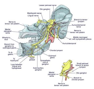
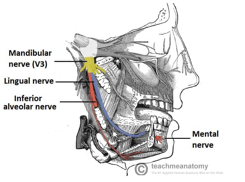
Anatomy, Head and Neck, Mandibular Nerve The fifth cranial nerve, the trigeminal nerve, has three branches which are the ophthalmic, maxillary, and mandibular. The third branch is called mandibular nerve (V3). It is the largest of the three divisions and carries both afferent and efferent fibers. The first two branches of the trigeminal ne …
Mandibular nerve (CN V3): Anatomy and course - Kenhub The mandibular nerve originates from the trigeminal ganglion of Gasser and exits the skull through the foramen ovale. Once it reaches the viscerocranium, it divides into two divisions: anterior and posterior. Both divisions further divide into smaller branches that innervate the structures of the face.
The Trigeminal Nerve: Anatomy, Function, and Treatment The small sensory branches of the trigeminal nerve have sensory endings located throughout the face, eyes, ears, nose, mouth, and chin. The branches of the trigeminal nerves travel along the pathways listed below. Ophthalmic The frontal nerve, the lacrimal nerve, and the nasociliary nerves converge in the ophthalmic nerve.
Trigeminal nerve - Wikipedia The three major branches of the trigeminal nerve—the ophthalmic nerve (V 1 ), the maxillary nerve (V 2) and the mandibular nerve (V 3 )—converge on the trigeminal ganglion (also called the semilunar ganglion or gasserian ganglion), located within Meckel's cave and containing the cell bodies of incoming sensory-nerve fibers.
Trigeminal nerve | Radiology Reference Article ... Although the trigeminal nerve is usually described singularly, it actually emerges/enters from the brain stem as multiple nerve roots 8. The sensory root is large and single and enters the anterolateral aspect of the pons. Usually superomedial to, and within 4 mm of the sensory root, one or more motor roots emerge.
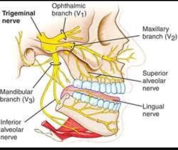
Trigeminal nerve anatomy, branches, distribution, function ... Trigeminal nerve. The large trigeminal nerve or 5th cranial nerve has three branches: ophthalmic (V1), maxillary (V2), and mandibular (V3) divisions. Trigeminal nerve is a mixed nerve providing sensations of the face for touch, temperature, and pain from the upper, middle, and lower portions of the face, as well as the oral cavity, to the brain.
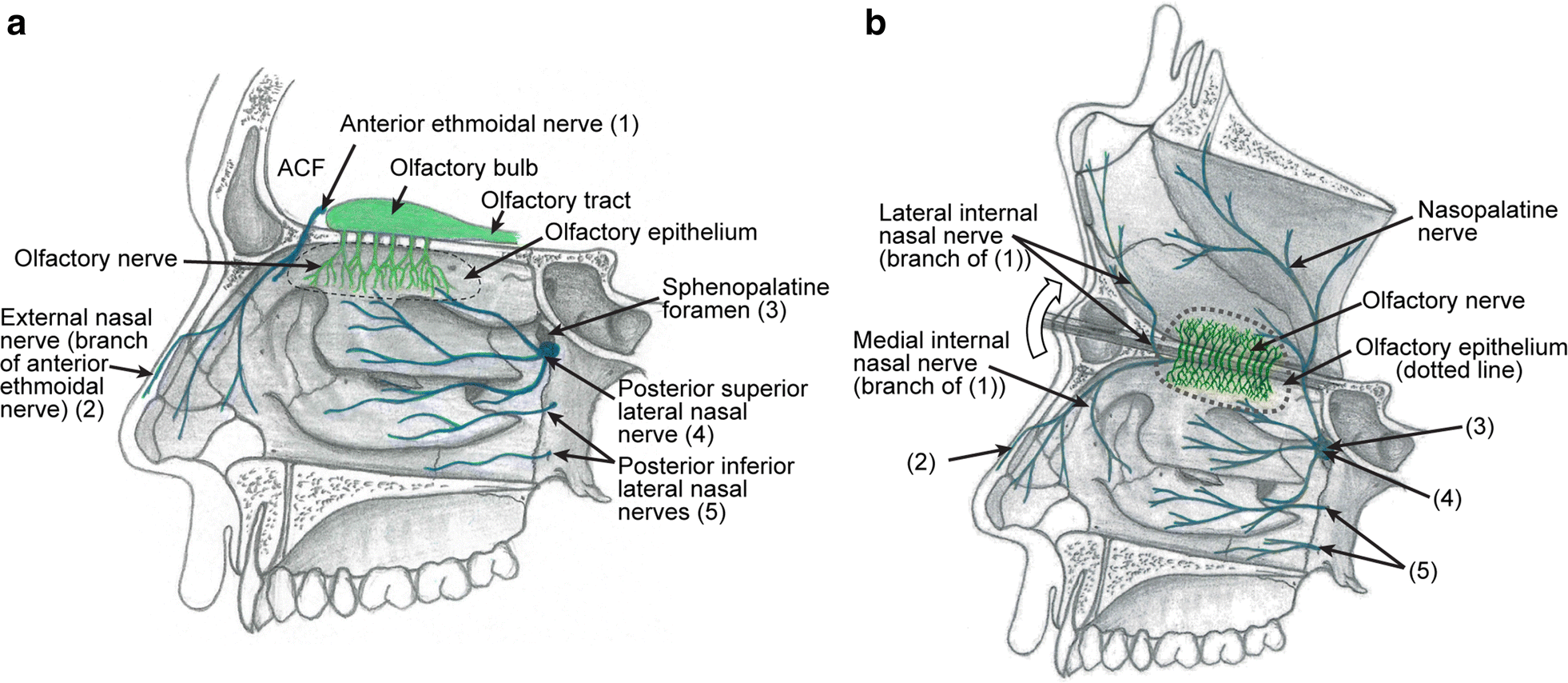
Trigeminal nerve route of entry. (A) Schematic showing the ... (A) Schematic showing the three branches of the trigeminal nerve: V1, V2, and V3. Branches V1 and V2 innervate the nasal cavity and project to the brain stem (BS).
Trigeminal nerve (CN V): Anatomy, function and branches ... The branches of the ophthalmic division of the trigeminal nerve are summarized below. Learn the anatomy of ophthalmic nerve with our videos, quizzes, articles, and labeled diagrams: Ophthalmic nerve (CN V1) Explore study unit Maxillary division (CN V2)
The Trigeminal Nerve (CN V) | Cranial Nerves | Geeky Medics Cranial nerves and cranial foramina diagram. 1. Embryology. The name trigeminal is derived from the Latin "tri-" meaning three, and "-geminal" meaning a group attached to a common point. There are 'three' major branches of the trigeminal nerve, coming from four distinct nuclei in the brainstem.
Trigeminal Nerve Anatomy Diagram shows trigeminal nerve (TGN), trigeminal ganglion, and peripheral divisions and their branches. From fora- men rotundum ossis sphenoidalis, maxil- lary nerve (thin underline) gains access to pterygopalatine fossa and continues in floor of orbit as infraorbital nerve. Inferior alveolar and lingual nerves ( thick underline )




:watermark(/images/watermark_5000_10percent.png,0,0,0):watermark(/images/logo_url.png,-10,-10,0):format(jpeg)/images/overview_image/274/CKsWYOqMbRE3da7XY9Ssw_the-maxillary-nerve_english.jpg)







:watermark(/images/watermark_5000_10percent.png,0,0,0):watermark(/images/logo_url.png,-10,-10,0):format(jpeg)/images/overview_image/522/hefybIAztz3WDIDI33S06g_the-mandibular-nerve_english.jpg)



0 Response to "36 trigeminal nerve branches diagram"
Post a Comment