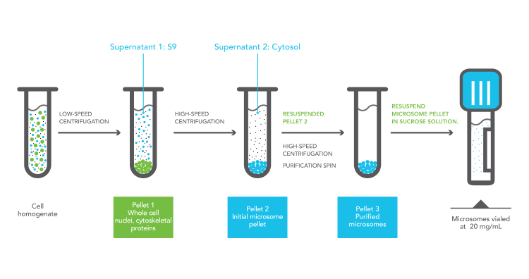
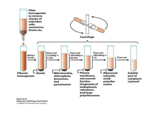
37 a diagram of cell fractionation is shown
O-6-methylguanine-DNA methyltransferase - Wikipedia O 6-alkylguanine DNA alkyltransferase (also known as AGT, MGMT or AGAT) is a protein that in humans is encoded by the O 6-methylguanine DNA methyltransferase (MGMT) gene. O 6-methylguanine DNA methyltransferase is crucial for genome stability.It repairs the naturally occurring mutagenic DNA lesion O 6-methylguanine back to guanine and prevents mismatch … Question paper (A-level) : Paper 3 - June 2018 - AQA small amount of cytoplasm and a cell-surface membrane. This very small daughter cell is called a polar body. Polar bodies do not usually divide. The same process occurs in the second division of meiosis, resulting in one egg cell and two polar bodies. The diagram in Figure 3 shows the formation of an egg cell and two polar bodies during meiosis ...
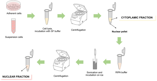
› articles › s41422/021/00530-9A phosphatidic acid-binding lncRNA SNHG9 facilitates LATS1 ... Jul 15, 2021 · As shown in Fig. 4j–l, small ... a Schematic diagram of subcellular fractionation procedures. ... Cell fractionation was conducted using Subcellular Protein Fractionation Kit (Pierce).

A diagram of cell fractionation is shown
› cell-biologyTop 16 Techniques Used in Cell Biology (With Diagram) ADVERTISEMENTS: The following points highlight the top sixteen techniques used in cell biology. Some of the techniques are: 1. Immunofluorescence Microscopy 2. Ion-Exchange Chromatography 3. Affinity Chromatography 4. Partition and Adsorption Chromatography 5. Gel Filtration Chromatography 6. Radioactive Tracer Technique 7. Radioimmunoassay (RIA) 8. Enzyme Immunoassay 9. Spectroscopy and ... › pmc › articlesMicroscale to Manufacturing Scale-up of Cell-Free Cytokine ... Feb 17, 2011 · a: Process flow diagram for protein purification (b) Cell-free reaction product pool was visualized, using non-reducing SDS–PAGE, by 14 C autoradiography of leucine incorporation at 5 h that measures only protein produced, or by Sypro staining of all proteins in the extract at 10 h. Actin Structure and Function - PubMed Central (PMC) 09.06.2011 · Shown are five actin molecules labeled –2 to +2. The run of the protein chain is shown as a secondary structure cartoon color-coded from blue (N terminus) to red (C terminus). Interacting side chains are shown as sticks. Panels b, d, and f are stereo pairs. Panels a and b show the main longitudinal interface between molecules 0 and 2. Panels c and d show the …
A diagram of cell fractionation is shown. (PDF) ESSENTIAL CELL BIOLOGY ESSENTIAL CELL BIOLOGY ... ESSENTIAL CELL BIOLOGY ESSENTIAL CELL BIOLOGY. × Close Log In. Log in with Facebook Log in with Google. or. Email. Password. Remember me on this computer. or reset password. Enter the email address you signed up with and we'll email you a reset link. Need an account? Click here to sign up. Log In Sign Up. Log In; Sign Up; more ... › doc › 123745376Biochemistry PDF | PDF | Cell (Biology) | Biochemistry Diagram of a typical prokaryotic cell. Enclosing the cell is the cell envelope generally consisting of a cell wall covering a plasma membrane though some bacteria also have a further covering layer called a capsule. The envelope gives rigidity to the cell and separates the interior of the cell from its environment, serving as a protective filter. Reassessment of Exosome Composition - PMC Apr 04, 2019 · (A) High-resolution iodixanol gradient fractionation of DiFi cell-derived (top) and primary human renal epithelial cell-derived (bottom) crude sEVs (P120) analyzed by immunoblotting. NV, non-vesicular; sEV, small extracellular vesicle. (B) Schematic of experimental design for Figure 6B – C. P120 sample was resupended and either loaded ... A single-cell transcriptomic landscape of the lungs of ... Dec 07, 2021 · The proportion of DEGs shared by the ageing and COVID-19 groups are shown for the different cell types. ... endothelial cell subtypes. j, Diagram of the coagulation ... fractionation for LC–MS ...
The Secretory Pathway: Randy Schekman - iBiology 00:09:14.23 And a Venn diagram of that distribution is shown in the next slide. 00:09:20.01 Most of the RNAs, most of the microRNAs, are found either entirely inside 293 cells, 00:09:28.18 or both in exosomes and in the cells. 00:09:32.21 Some 90% of the RNAs are not enriched in the extracellular vesicles. (PDF) Molecular Cell Biology 5th ed - Lodish et al | 희수 김 ... Molecular Cell Biology 5th ed - Lodish et al. × Close Log In. Log in with Facebook Log in with Google. or. Email. Password. Remember me on this computer. or reset password. Enter the email address you signed up with and we'll email you a reset link. Need an account? Click here to sign up. Log In Sign Up. Log In; Sign Up ... Actin Structure and Function - PubMed Central (PMC) 09.06.2011 · Shown are five actin molecules labeled –2 to +2. The run of the protein chain is shown as a secondary structure cartoon color-coded from blue (N terminus) to red (C terminus). Interacting side chains are shown as sticks. Panels b, d, and f are stereo pairs. Panels a and b show the main longitudinal interface between molecules 0 and 2. Panels c and d show the … › pmc › articlesMicroscale to Manufacturing Scale-up of Cell-Free Cytokine ... Feb 17, 2011 · a: Process flow diagram for protein purification (b) Cell-free reaction product pool was visualized, using non-reducing SDS–PAGE, by 14 C autoradiography of leucine incorporation at 5 h that measures only protein produced, or by Sypro staining of all proteins in the extract at 10 h.
› cell-biologyTop 16 Techniques Used in Cell Biology (With Diagram) ADVERTISEMENTS: The following points highlight the top sixteen techniques used in cell biology. Some of the techniques are: 1. Immunofluorescence Microscopy 2. Ion-Exchange Chromatography 3. Affinity Chromatography 4. Partition and Adsorption Chromatography 5. Gel Filtration Chromatography 6. Radioactive Tracer Technique 7. Radioimmunoassay (RIA) 8. Enzyme Immunoassay 9. Spectroscopy and ...
A protocol for the subcellular fractionation of Saccharomyces ...

A reproducible method for the extraction, identification and ...

Organization of the Cell - ppt video online download

Herpes simplex virus 1 α regulatory protein ICP0 functionally ...
Results for "Cell Fractionation" | Springer Nature Experiments

Functional Cell Surface Display and Controlled Secretion of ...
LI: To understand what cell fractionation is and be able to ...
Improvement of Electrochromic Performance by Embedding ITO ...

Cellular organelles and structure (article) | Khan Academy

Copyright © 2005 Pearson Education, Inc. publishing as ...
Fractionation of cells

cell_fractionation - YouTube
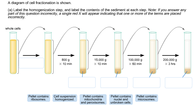
Solved A diagram of cell fractionation is shown (a) Label ...

Sequential fractionation and isolation of subcellular ...

Cell Fractionation
Solved A diagram of cell fractionation is shown (a) Label ...

How to Prepare Protein Samples for Western Blot | GoldBio

Experiment 1 - CellBiologyOLM
Cell fractionation and ultracentrifugation AS level ...

Cell Fractionation: Definition, Steps & Methods - Video ...
Frontiers | High Resolution Proteomic Analysis of Subcellular ...

Subcellular Proteomics” of Neuromelanin Granules Isolated ...
PLOS ONE: Proteome analysis of Phytomonas serpens, a ...

IJMS | Free Full-Text | Interaction of Alpha Synuclein and ...
Frontiers | Cellular Fractionation and Nanoscopic X-Ray ...
Subcellular Fractions | BioIVT
Optimization of a multi-well colorimetric assay to determine ...
Merging high-quality biochemical fractionation with a refined ...

Phosphate and phosphite differentially impact the proteome ...

Subcellular Fractionation

Fractionation - an overview | ScienceDirect Topics

Cell Fractionation - an overview | ScienceDirect Topics
Cell Organelle Fractionation
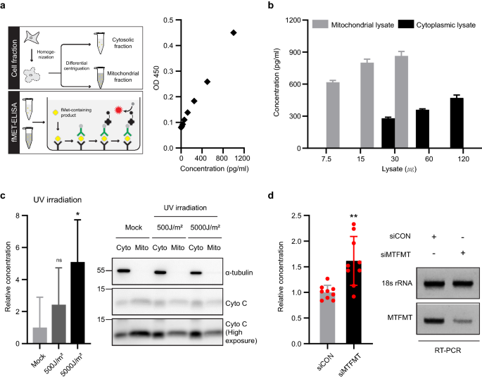
MTFMT deficiency correlates with reduced mitochondrial ...
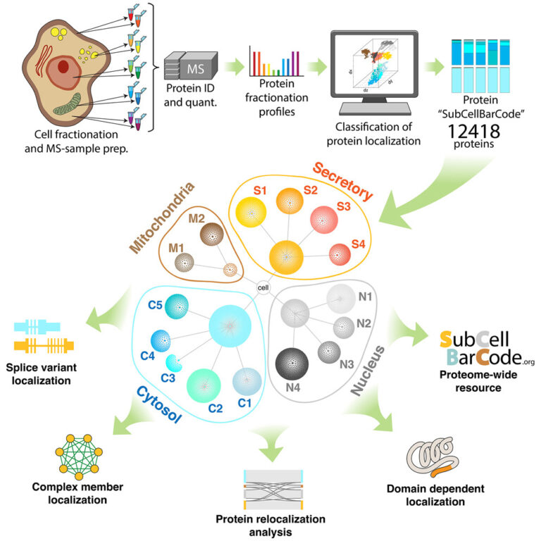
New analysis method for mapping proteins shared in an open ...
Cell Organelle Fractionation

786-249
0 Response to "37 a diagram of cell fractionation is shown"
Post a Comment