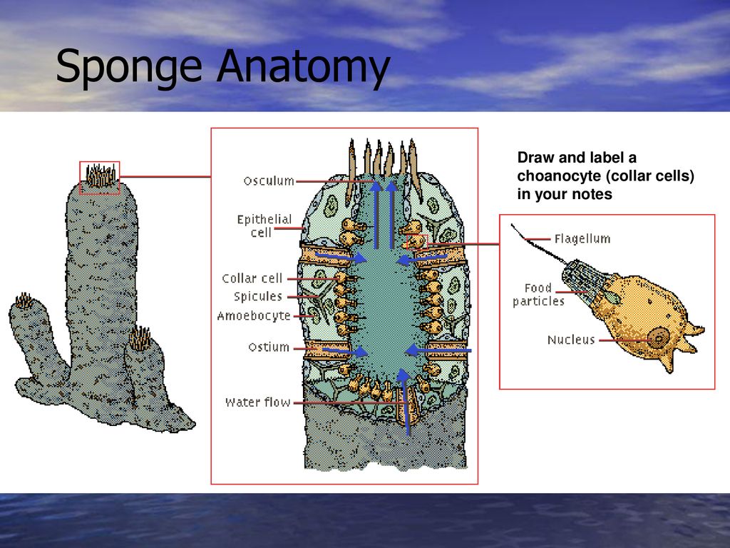
40 sponge diagram with labels
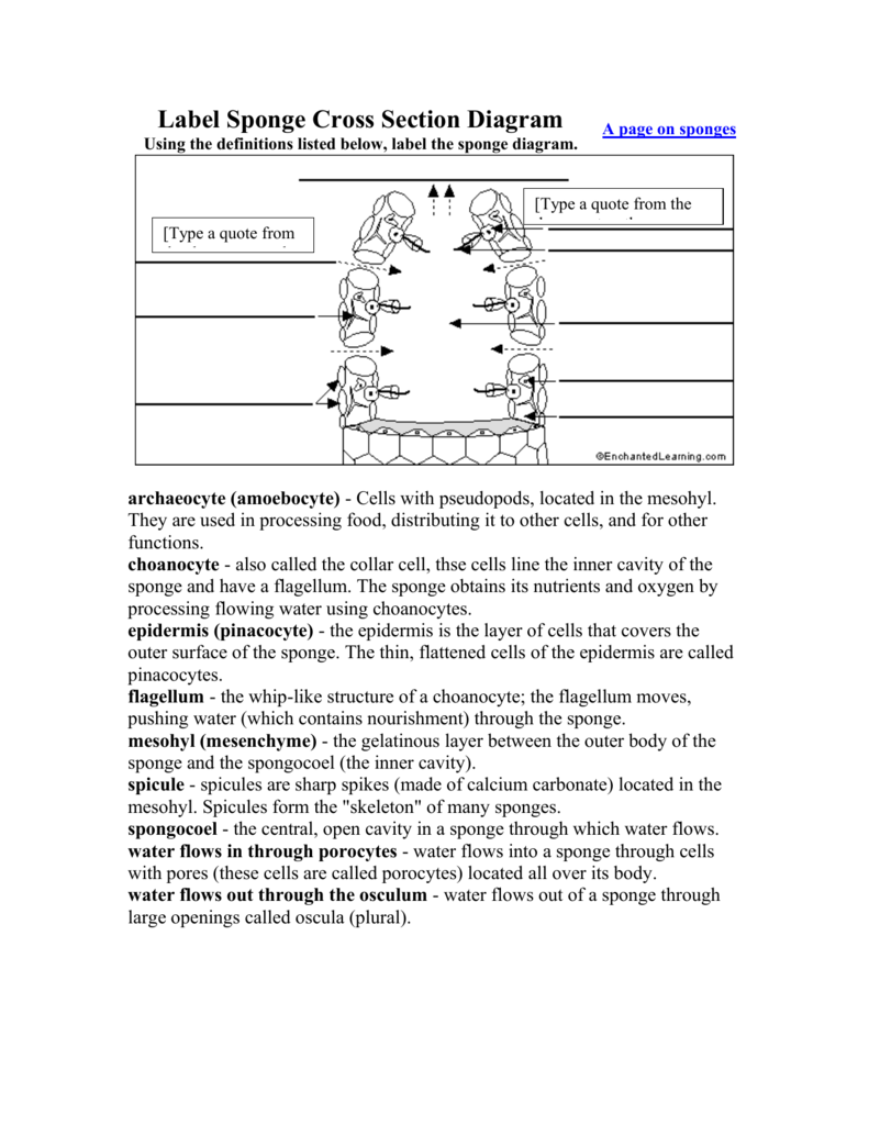
Label Sponge Cross Section Diagram Using the definitions listed below, label the sponge diagram. A page on sponges archaeocyte (amoebocyte) - Cells with pseudopods, located in the mesohyl. They are used in processing food, distributing it to other cells, and for other functions. Sponge Anatomy Labeling Page. Higher Resolution PDF for Printing. Click Here. Link to More Info About this Animal (with Labeled Body Diagram) Click Here. Citing Research References. When you research information you must cite the reference. Citing for websites is different from citing from books, magazines and periodicals. The style of citing ...
The glomerulus is a ball of capillaries surrounded by the Bowman's capsule into which urine is filtered. The filtration barrier consists of 3 components: Endothelial cells of glomerular capillaries Glomerular basement membrane Epithelial cells of Bowman's Capsule (podocytes) This article shall consider the structure of the filtration barrier, filtration and relevant clinical conditions.

Sponge diagram with labels
Sponge Anatomy Coloring Page. PDF for Printing Out. Click Here. Link to More Info About this Animal (with Labeled Body Diagram) Click Here. Citing Research References. When you research information you must cite the reference. Citing for websites is different from citing from books, magazines and periodicals. The style of citing shown here is ... 1. The vulva. It's a common misconception that the visible outer parts of the female anatomy is called the vagina. The technical name is actually the vulva. Yours has the job of protecting your ... 1.A) Discuss the life cycle of the jellyfish Aurelia. Is there an Alternation of Generations in the life cycle? Explain. 1.B) 11. How does the life cycle of Obelia differ from the life cycle of Hydra? What is the name of the feeding polyp of Obelia? The reproductive polyp?
Sponge diagram with labels. Biotin-labeled miR-370-5P and negative control probes were synthesized by RiboBio (Guangzhou, China). Anti-Ago2 RNA-binding protein immuno-precipitation (RIP) assay Dual luciferase assay The PS-Spot refers to the perineal sponge. It's a spongy cushion of tissue and blood. It sits between the vaginal opening and rectum and is located internally to the perineum (which is the name for the area between vagina and rectum). The U-Spot. The U-Spot is the sensitive area between the clitoris and the urethra. The male reproductive system consists of many different parts and structures which are all equally important. The task of the reproductive system in men is to produce male sex hormones and to make, preserve and carry the sperm as well as the semen. It is responsible for discharging sperm into the female reproductive tract and fertilizing an egg. Animal Cell Diagram - Labeled - Tim's Printables from www.timvandevall.com Oct 04, 2019 · ncert solutions for class 8 science chapter 8 cell structure and functions topics and sub topics in class 8 science chapter 8 cell structure and functions: Active and passive transport, brief explanation of facilitated diffusion (uniport, symport and ...
Project Network Diagram Template Sep 27, 2019 · A network diagram will help organizations and teams visualize how devices like computers, and networks like telecommunications, work together. Network diagrams help paint a picture of how these operational networks function and they identify components like routers, firewalls and devices, and ... Label and diagram the difference. Horseshoe Crab Printable - A horseshoe crab is an armored hard shell crab. Learn about their diet and diagram their body parts. Lesson 9: Mollusks. Clam Printable - Diagram and learn about the clam with this printable. Oyster Labeling Sheet - Label the different parts of the oyster and learn about pearls. The diagram shows a sponge. What is the function of the structures labeled X? providing protection from predators fertilizing eggs filtering and digesting food stinging prey DO NOT ANSWER WITH A VIRUS LINK!!!!!~!!!!!11!!!!!1!1!!!!! 1 See answer gamerplayz845 is waiting for your help. ... The diagram shows a sponge. - 22494222 gamerplayz845 gamerplayz845 03/24/2021 Biology Middle School answered The diagram shows a sponge. What is the function of the structures labeled X? providing protection from predators fertilizing eggs filtering and digesting food stinging prey 2 See answers Advertisement Advertisement sophiayarushina123 ...
Mesophyll cells are found in the plant's leaves. They are a type of ground tissue that is actually found as two distinct types in the leaves. In a nice organized order we find the palisade ... Bone histology. The strength, shape and stability of the human body are dependent on the musculoskeletal system. The most robust aspect of this unit is the underlying bony architecture. Bone is a modified form of connective tissue which is made of extracellular matrix, cells and fibers. Sponge-dwelling fauna: a review of known species from the Northwest Tropical Atlantic coral reefs ... We also created an interactive visualisation of the species-interactions dataset and of a dynamic Chord Diagram of the host-guest species connections to generate a user-friendly link between the user and the dataset. ... Column label Column ... Draw And Label Sponges / Having observed the sponge anatomy, draw a simple sketch of a sponge and label the following parts: Beating of choanocyte flagella draws water through the sponge so that . Spongin, spicules, ostia, choanocytes, osculum. Sponge...

Porifea - Skeletal System
Sponges. The Sponges ClipArt gallery includes 48 illustrations of sponges. Sponges, also called poriferans, are in the phylum porifera and are all sessile animals that live and feed attached to the bottom of the sea.

Science Chapter 30 Sponges and Cnidarians 🧽 🦑 Diagram | Quizlet
"Diagrams to illustrate the development of one of the simpler types of sponge: I, the egg; 2, section… Spongilla "Vertical section of a fresh-water sponge (Spongilla), showing the arrangement of the canal-system.…
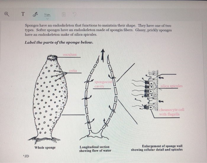
Solved T & Sign Sponges have an endoskeleton that functions ...
Furthermore, a 142.5-fold enriched miR-24-3p was captured with the biotin-labeled circRtn4 in RAP assay (Figures 5D,E), which revealed that circRtn4 could act as a sponge of miR-24-3p. Open in a separate window
Vector Illustration Marine Sponge Marine Sponge Stock Vector ...
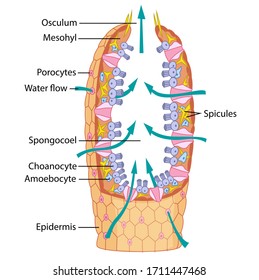
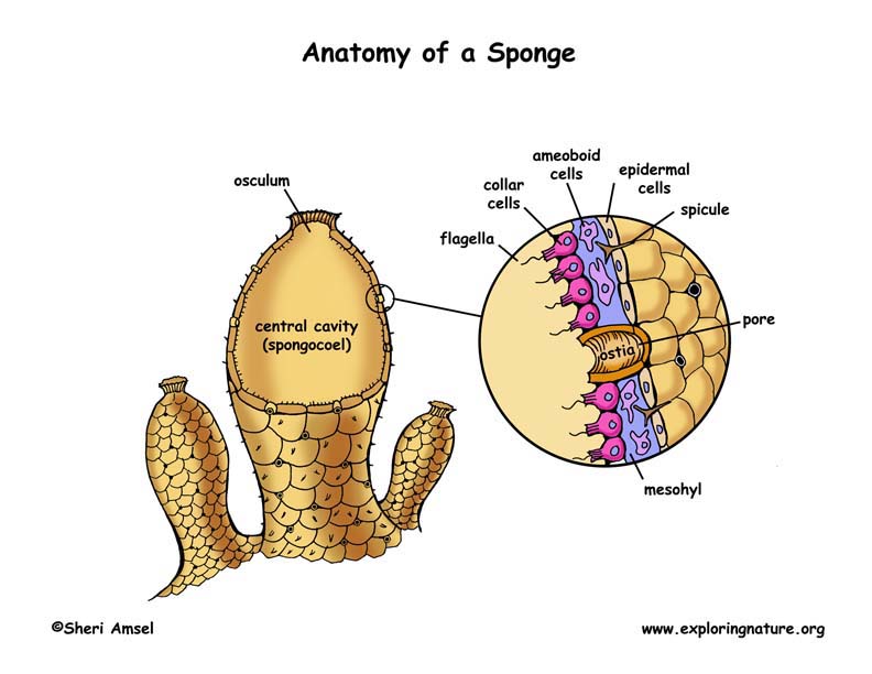
Label a diagram of a sponge (including the spongocoel, choanocyte, mesohyl, amoebocyte, osculum, and spicules). Describe the function of each part. 3. Describe how a sponge feeds and digests its food. Cnidaria 4. List the characteristics of the phylum Cnidaria that distinguish it from other animal phyla. 5.
Reading: Sponges | Biology II Laboratory Manual
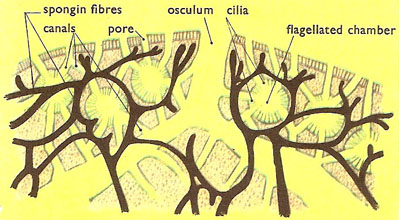
The following diagrams come from Invertebrate Zoology, by Rupert and Barnes. The photos are my own. Here is a close view of an asconoid sponge from our aquaria in the lab - similar if not identical to the one in Fig 5-3 B above:

BIO1403 - EXAM 1 (SPONGES) Diagram | Quizlet
Label Sponge External Anatomy Diagram Using the definitions listed below, label the sponge and the flow of water through it. A page on sponges epidermis - the layer of cells that covers the outer surface of the sponge. The thin, flattened cells of the epidermis are called pinacocytes. holdfast - root-like tendrils that attach the sponge to rocks.
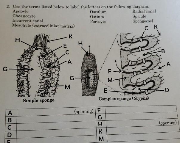
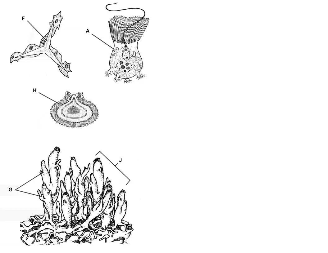
Solved 2. Use the terms listed below to label the letters on ...
Sponge Function Diagram SHA-3's sponge construction works by: Breaking the input data to be hashed into r-bit sized chunks, in SHA-3's case the rate+capacity bits sum up to 1600 bits. The first input rate and capacity are all zeros and are XORed with the first rate block r 1

Phylum Porifera The Sponges | Ekohadiprabowo's Blog
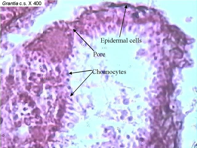
Choanocytes have a round cell body that's attached to the inside wall of the sponge and is also the location of the cell's nucleus and food vacuoles. Each choanocyte has a single flagellum , which ...
Phylum Porifera The Sponge. - ppt download
39 make an electron distribution diagram of water. Solution for Draw the electron distribution diagram for water. Begin with 1 central water molecule. Show the chemistry of each element within the central ... Make an electron distribution diagram of water. Yahoo Answers has shut down as of May 4, 2021. Yahoo Answers was once a key part of Yahoo ...
What's a Tunicate?
Outlines the distinguishing features of the Phlyum Porifera with a labelled diagram of a sponge. They are bottom-dwelling fish living down to 200 m although they can be found in much shallower water. Lower classifications of sea stars include Brisingida Velatida Spinulosida.

Pin on Phylum Porifera
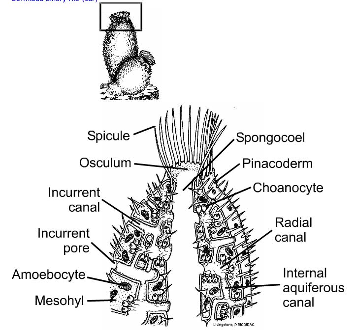
A sponge endoskeleton consists of short, sharp rods called spicules (see Figure below). Spicules are made of silica, calcium carbonate, or spongin, a tough protein. They grow from specialized cells in the body of the sponge. Sponge Anatomy. A sponge lacks tissues and organs, but it has several types of specialized cells. Sponges are filter feeders.
Label Sponge External Anatomy - EnchantedLearning.com
Remove any stickers or labels from the appliance. Thoroughly clean the accessories with hot water: use some mild detergent and a non-abrasive sponge. Wipe the inside and outside of the appliance with a damp cloth. initial start-up operation may produce a smell and smoke (For about 15 mins.). This is normal.
Porifera
Fig: Labeled Diagram of Sponge. Learn Exam Concepts on Embibe. 8. The nervous and sensory cells are probably not differentiated. 9. Canal System - It is also known as the Aquiferous system, and it is a system of interconnected canals through which water circulates and helps in a number of metabolic activities of a sedentary sponge.
sponge
Porifera (Sponge) Quiz. . 1. What is the name of these Porifera structures? 2. What is the name of cells that generate a flow of water through the sponge? 3. Choanocyte cells create a current that bring in O2 and small molecules and carry away CO2 and NH3. What part of a Coanocyte sets up a water current?

Sponge | Plant science, Sponge, Marine biology
Porifera Temporal range: Ediacaran-recent PreꞒ Ꞓ O S D C P T J K Pg N A stove-pipe sponge Scientific classification Kingdom: Animalia Phylum: Porifera Grant, 1836 Classes Calcarea Hexactinellida Demospongiae Homoscleromorpha † Stromatoporoidea † Archaeocyatha Synonyms Parazoa /Ahistozoa (sans Placozoa) Sponges, the members of the phylum Porifera (meaning 'pore bearer'), are a basal ...

Sponge Labeling Diagram (1).pdf - Sponge Labeling Diagram ...
The Main Parts of a Dishwasher (Diagram Included) Steve Updated June 15, 2021 May 17, 2021. According to the U.S. Energy Information Administration (EIA), approximately 80 million households in the United States have a dishwasher. If you have one, there is a good chance that this is the appliance that routinely saves you the most time.
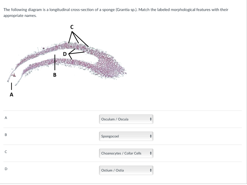
Solved The following diagram is a longitudinal cross-section ...
1.A) Discuss the life cycle of the jellyfish Aurelia. Is there an Alternation of Generations in the life cycle? Explain. 1.B) 11. How does the life cycle of Obelia differ from the life cycle of Hydra? What is the name of the feeding polyp of Obelia? The reproductive polyp?
Sponge Coloring Diagram and Questions - BIOLOGY JUNCTION
1. The vulva. It's a common misconception that the visible outer parts of the female anatomy is called the vagina. The technical name is actually the vulva. Yours has the job of protecting your ...

Porifera - Kingdom AnIMALIA
Sponge Anatomy Coloring Page. PDF for Printing Out. Click Here. Link to More Info About this Animal (with Labeled Body Diagram) Click Here. Citing Research References. When you research information you must cite the reference. Citing for websites is different from citing from books, magazines and periodicals. The style of citing shown here is ...
Label Sponge Cross-Section - EnchantedLearning.com

Label the Sponge Diagram | Quizlet
Phylum - Porifera (Sponges)
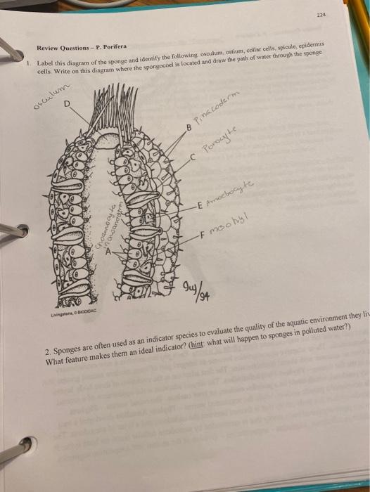
Solved 224 Review Questions - P. Porifera Label this diagram ...

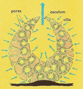
3. Diagrammatic representation of a simple (asconoid) sponge ...

Sponge Labeling and Sponge Review Diagram | Quizlet

Types of locally labeled sea sponges. a: drawing of a vase ...
Sponges - EnchantedLearning.com
sponge
Biology 10: Topic 10a: Simple Invertebrates - Sponges

Sponge | Biology plants, Study notes, Life science

Introduction to Sponges Porifera Porifera Porebearing Over 4
Label Sponge Cross Section Diagram_Blank

Sycon diagram with Detailed Illustrations and Clear Labels

Sponge Structure and Function - Advanced ( Read ) | Biology ...

Biology Ch. 33- Label the Parts of a Sponge Diagram | Quizlet

Sponge Printout - EnchantedLearning.com

sponges/ phylum porifera Diagram | Quizlet

Modern sponge anatomy. ( A ) Schematic cross-section of ...
Overview of Sponges

Porifera : Characteristics and Classification - Biology Edu Care
0 Response to "40 sponge diagram with labels"
Post a Comment