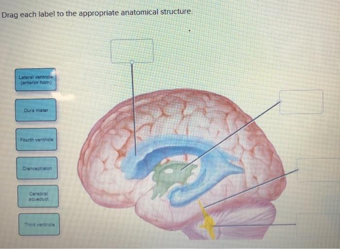
38 drag the labels onto the diagram of the cns meninges.
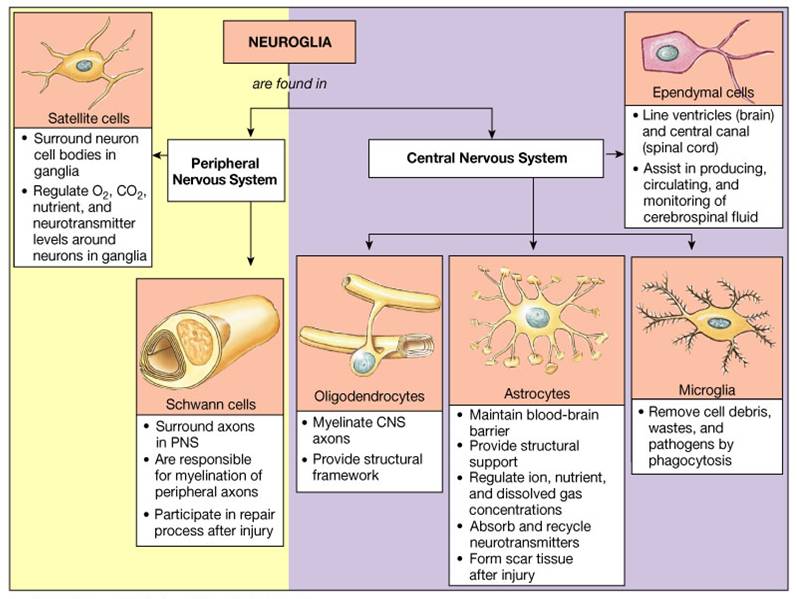
A&P2 Lab 3 HW Flashcards | Quizlet Terms in this set (15) Drag the labels onto the diagram to identify the gross anatomical structures of the spinal cord. Drag the labels onto the diagram to identify the spinal nerve roots and meninges. Drag the labels onto the diagram to identify the parts of the spinal cord (transverse section, showing white matter). A&P2 Lab and Powerpoints Flashcards - Quizlet Drag the labels onto the diagram to identify the gross anatomical structures of the spinal cord. look at pic. Drag the labels onto the diagram to identify the spinal nerve roots and meninges. look at pic. ... while Central Nervous System (CNS) neuroglial cells called _____ are responsible for the formation of a myelin sheath. Schwaan cells ...
Spinal Cord Quiz: Cross-Sectional Anatomy - GetBodySmart An interactive quiz covering Spinal Cord Cross-Sectional Anatomy through multiple-choice questions and featuring the iconic GBS illustrations.
Drag the labels onto the diagram of the cns meninges.
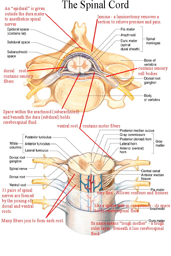
Week 4 Chapter 13_.pdf - Week 4 Chapter 13 ... - Course Hero Part A Drag the labels to the appropriate location in the figure. ANSWER: control of the three trimesters of pregnancy regulation of the multiyear puberty process release of insulin after a meal detection of pain after a bee sting A neuron on the left side of the spinal cord sends an impulse over to the right side of the spinal cord. Solved Part A Drag the labels onto the diagram to identify ... Question: Part A Drag the labels onto the diagram to identify the spinal nerve roots and meninges Reset Help Ventral Pia mater Meninges Dorsal root Dura mater . This problem has been solved! See the answer See the answer See the answer done loading. Show transcribed image text Expert Answer. Spinal Cord - Anatomy, Structure, Function, & Diagram Spinal Cord Anatomy. In adults, the spinal cord is usually 40cm long and 2cm wide. It forms a vital link between the brain and the body. The spinal cord is divided into five different parts. Several spinal nerves emerge out of each segment of the spinal cord. There are 8 pairs of cervical, 5 lumbar, 12 thoracics, 5 sacral and 1 coccygeal pair ...
Drag the labels onto the diagram of the cns meninges.. Sheep Brain Dissection with Labeled Images Brain with Dura Mater Intact. Removal of the Dura Mater. 2. This image shows the ventral surface of the sheep's brain with most of the dura mater removed. The pituitary gland and the optic chiasma are still intact. (A = pituitary gland, B = optic chiasma, C = olfactory bulb) 3. On this image, the dura matter has been completely removed, you can ... Diagram Of Eye Visual Axis Medial Lateral - Studying Diagrams Drag the labels onto the diagram to identify the sel anatomy of the eye supio view Reset Help Visual axis Posterior Anterior Edge of chamber chambepupi Ciliary. LATERAL GENICULATE NUCLEUS OF THE SHEEP O 312-10h2 Largecells29 039 I I 1 mm I Fig. Download full-size image Figure 2. Brain Diagram Labeled Quizlet - Studying Diagrams Pin By Daniela Diaz On Anatomy Lab Brain Diagram Nervous System Anatomy Brain Anatomy In 2021 Brain Diagram Nervous System Anatomy Human Brain Diagram. Start studying Brain meninges labeled. This part of the cerebrum interprets and sorts information from the senses. Drag and drop the text labels onto the boxes next to the heart diagram. PDF DISSECTION OF THE SHEEP'S BRAIN - Hanover College Sheep Brain Dissection Guide Now onto the Dissection Proper. ... The meninges are the protective coverings, which enclose the brain and spinal cord. The dura mater, the tough outer layer, will have been mostly ... the central nervous system). This is the corpus callosum. It is so big that different parts of it get different names.
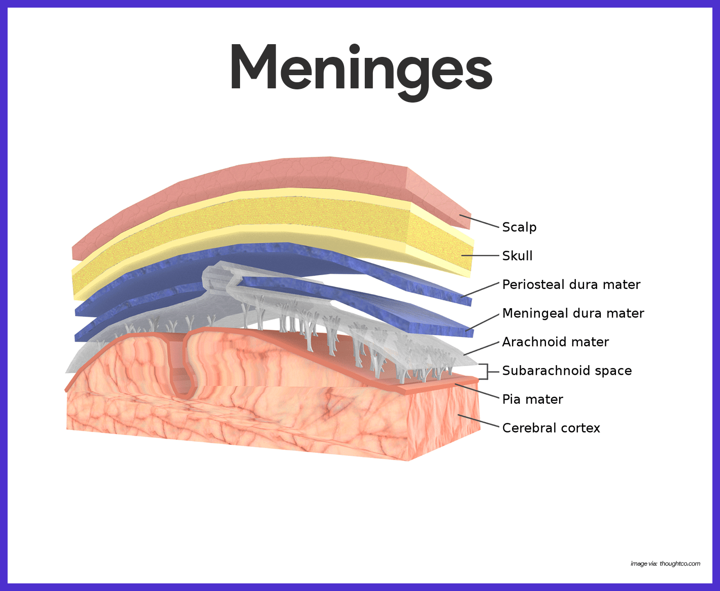
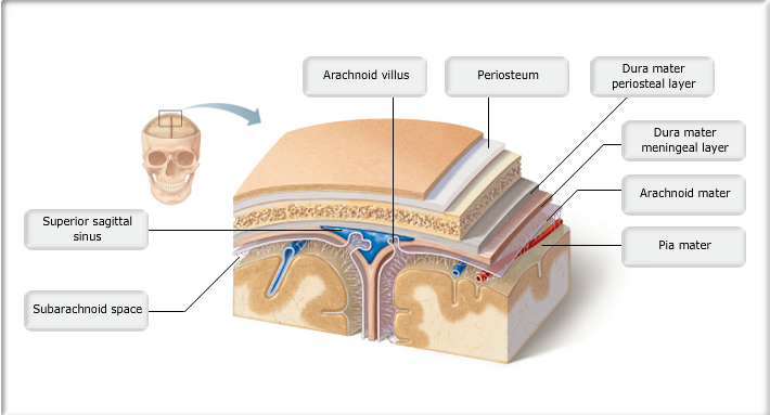
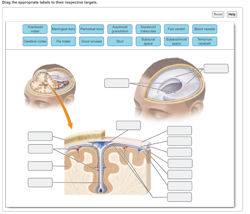
ingenieurbuero-edgar-pech.de Drag The Labels Onto The Diagram To Identify The Structures And Ligaments Of The Shoulder Joint. The bar button is labeled with the name of the symbol palette. Name the new process System. Select subVI you wish place 4. To enter text into diagram shapes May 05, 2010 · First add data labels to the chart (Layout Ribbon > Data Labels) Define the ... Ventricles of the Brain: Anatomy, Function, CSF Flow - EZmed Learn the ventricles of the brain along with their definition, function, location, anatomy, and cerebrospinal fluid (CSF) flow using labeled diagrams. The ventricular system contains the lateral, third, and fourth ventricles whose function is to produce cerebrospinal fluid. Learn where CSF is found, Major Body Cavities & their subdivisions Flashcards by ... Meninges The membranes covering both the brain and the spinal cord 5 Ventral body cavity The more anterior and large of the closed body cavities; houses internal organs collectively called the viscera, or visceral organs; has two major subdivision, the thoracic cavity and the abdominopelvic cavity ... Solved Part A Drag the labels onto the diagram to identify ... Question: Part A Drag the labels onto the diagram to identify the cranial meninges and associated structures. Reset Hel Subdural space Pia mater Subarachnoid space Dural sinus Cranium Dura mater (meningeal layer) Dura mater (periosteal layer) Arachnoid mater Cerebral cortex QUID Submit Request Answer.
CT Brain Anatomy - Calcified structures Key points. Commonly calcified structures of the brain include the choroid plexus, pineal gland, basal ganglia and falx. Use of CT 'bone windows' is helpful in differentiating calcified structures from acute haemorrhage. There are several structures in the brain which are considered normal if calcified. Knowledge of these structures helps avoid ... A&P2 Lab 2 HW Flashcards | Quizlet Drag the labels onto the diagram to identify the origins of the cranial nerves (VII - XII). look at pic The accumulation of blood during an epidural or subdural hemorrhage creates debilitating pressure on the brain and, without help, death is imminent. Where exactly is blood accumulating in a subdural hemorrhage? priessnitzumschlag.de To label an element, double-click the element and enter the label text in the text box. Drag a List box from the Controls stencil and position it in the upper-left corner of the panel container as shown in the following graphic. Start a sequence diagram. - Drag The Labels Onto The Diagram To Identify Structures . lab 7 (exercise 14) Flashcards - Quizlet Drag the labels onto the diagram to identify the cranial meninges and associated structures. 1. dura mater 2. subarachnoid space 3. pia mater 4. cerebral cortex 5. cranium 6. periosteal cranial dura 7. dural sinus 8. meningeal cranial dura 9. subdural space 10. arachnoid mater
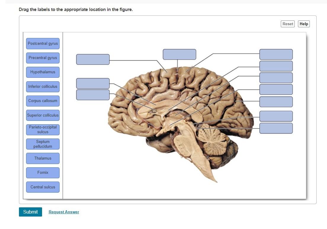
Solved Drag the labels to the appropriate location in the ...
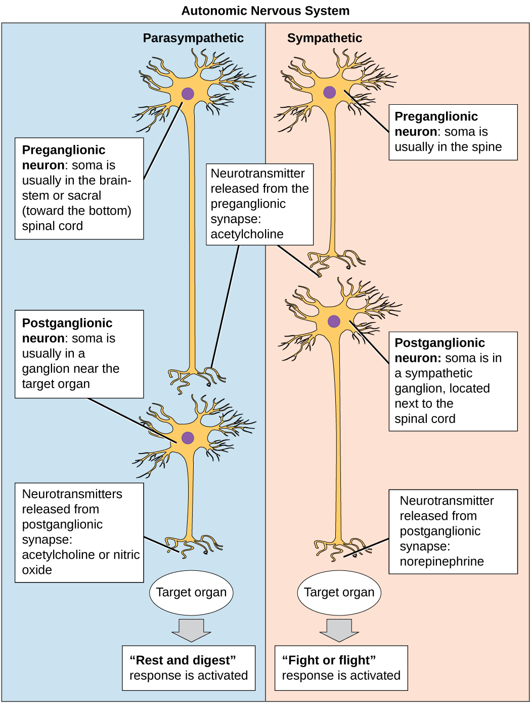
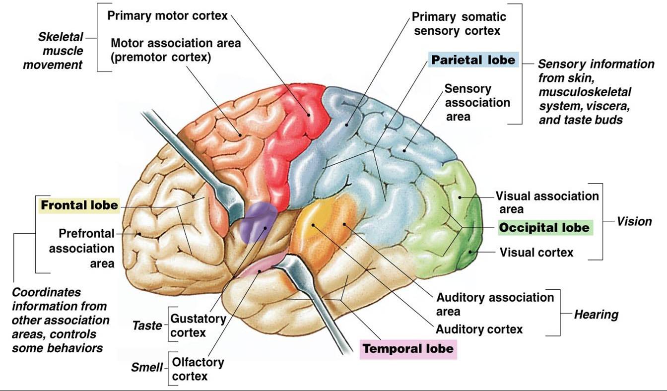
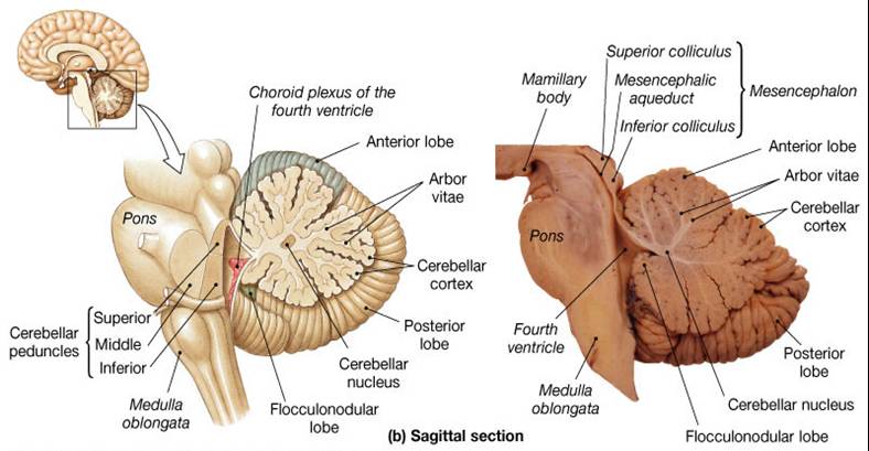
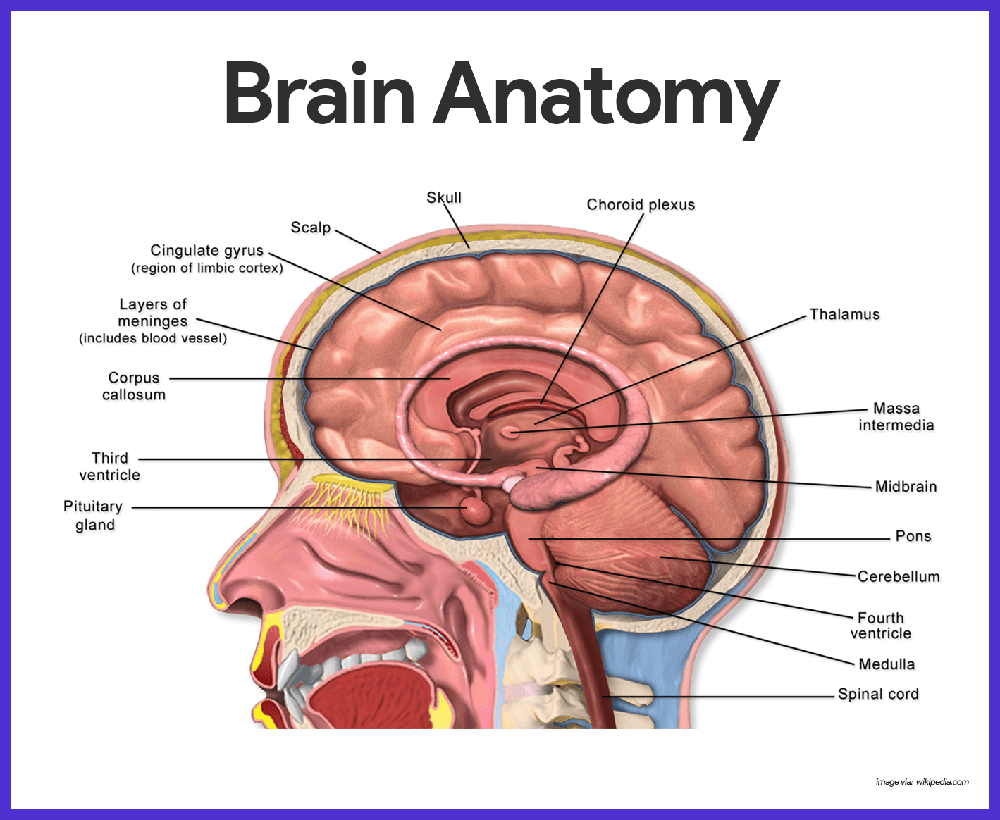
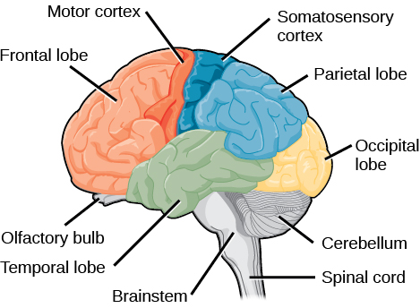
14.3 The Brain and Spinal Cord - Anatomy & Physiology The brain and the spinal cord are the central nervous system, and they represent the main organs of the nervous system. The spinal cord is a single structure, whereas the adult brain is described in terms of four major regions: the cerebrum, the diencephalon, the brain stem, and the cerebellum. A person's conscious experiences are based on ...
11.6 Nervous System – Concepts of Biology – 1st Canadian Edition
Lab Activity Chapter 17.pdf - 4/4/2020 Lab Activity ... Part A Drag the labels onto the diagram to identify the parts of the dissected sheep brain, median section (part 2 of 2). ANSWER: Correct Art-labeling Activity: Figure 17.4a (1 of 3) Part A Drag the appropriate labels to their respective targets.
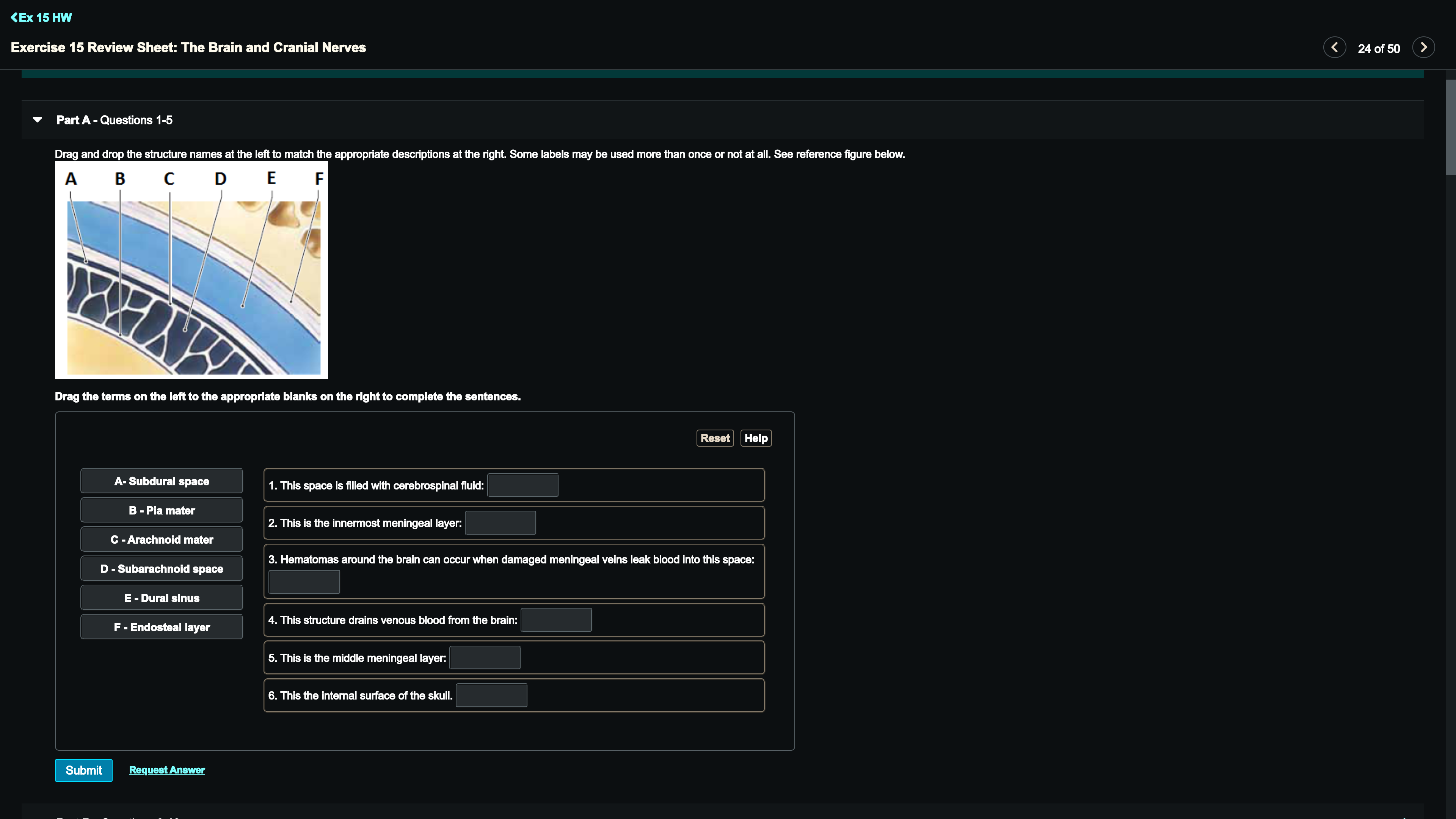
Solved Part A - Questions 1-5 Drag and drop the structure ...
Exercise 1 Language of Anatomy.pdf - Course Hero Part A Drag the labels onto the diagram to identify the regions of the appendages. ANSWER: Correct Exercise 1 Review Sheet Art-labeling Activity 1 (1 of 3) Identify the regional terms on an anterior view of the trunk. Part A Drag the labels onto the diagram to identify the regions of the trunk.
Nervous System Anatomy and Physiology - Nurseslabs
A&P2 Lab 13 HW, A&P2 Lab 12 HW, A&P2 Lab 11 HW ... - Quizlet Drag the labels onto the diagram to identify the cranial meninges and associated structures. look at pic Drag the labels to identify the landmarks and features on one of the cerebral hemispheres.

Case Study - Stroke A 77-year-old woman was cooking in the ...
cerebral cortex diagram The Cerebral Cortex The Cerebral Cortex diagram and chart - Human body anatomy diagrams and charts with labels. Visual cortex and other functional modalities mapped onto a surface-based atlas of macaque cerebral cortex. Functional Anatomy of Basal Ganglia Circuits with the ... Cerebral Cortex Stock Photos, Pictures & Royalty-Free ...

Solved Part A Drag the labels onto the diagram to identify ...
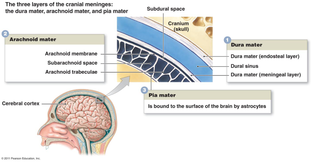
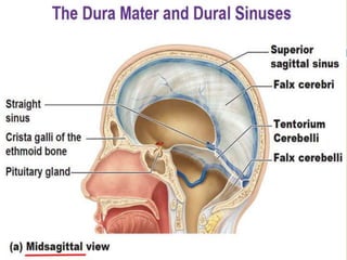
Neuroanatomy, Cranial Meninges - StatPearls - NCBI Bookshelf The brain and spinal cord are enveloped within three layers of membrane collectively known as the meninges, with the cranial meninges specifically referring to the section that covers the brain. From superficial to deep, the three layers are the dura, arachnoid, and pia—the term "mater," Latin for mother, often follows these names (i.e., dura mater, arachnoid mater, pia mater).[1]
11.6 Nervous System – Concepts of Biology – 1st Canadian Edition
Cranial nerves quizzes and labeling exercises - Kenhub Free labeling quiz. Try to understand and memorize what you can from the labeled diagram, then, try to label the cranial nerves yourself with our cranial nerves labeling quiz exercise available to download below. This is a great way to start to get the cogs turning and warm up your memory before you take our other cranial nerve quizzes (but one ...
Associate Degree Nursing Physiology Review
Screen Shot 2019-02-05 at 8.26.00 PM.png - Drag the labels ... View Screen Shot 2019-02-05 at 8.26.00 PM.png from BSC 2086L at University of South Florida. Drag the labels onto the diagram to identify the cranial meninges and associated structures.

14.2 Blood Flow the meninges and Cerebrospinal Fluid ...
Admissions and Records - ApplyOUHSC Mailing Address: P.O. Box 26901, LIB-121, Oklahoma City, OK 73126 Location: 1105 N. Stonewall Ave., Oklahoma City, OK 73117-1211 Telephone: (405) 271-2359 FAX: (405 ...
Associate Degree Nursing Physiology Review
Spinal Cord - Anatomy, Structure, Function, & Diagram Spinal Cord Anatomy. In adults, the spinal cord is usually 40cm long and 2cm wide. It forms a vital link between the brain and the body. The spinal cord is divided into five different parts. Several spinal nerves emerge out of each segment of the spinal cord. There are 8 pairs of cervical, 5 lumbar, 12 thoracics, 5 sacral and 1 coccygeal pair ...
BIOL 237 Class Notes - The Spinal Cord and Spinal Nerves
Solved Part A Drag the labels onto the diagram to identify ... Question: Part A Drag the labels onto the diagram to identify the spinal nerve roots and meninges Reset Help Ventral Pia mater Meninges Dorsal root Dura mater . This problem has been solved! See the answer See the answer See the answer done loading. Show transcribed image text Expert Answer.
Nervous System Anatomy and Physiology - Nurseslabs
Week 4 Chapter 13_.pdf - Week 4 Chapter 13 ... - Course Hero Part A Drag the labels to the appropriate location in the figure. ANSWER: control of the three trimesters of pregnancy regulation of the multiyear puberty process release of insulin after a meal detection of pain after a bee sting A neuron on the left side of the spinal cord sends an impulse over to the right side of the spinal cord.
Anatomy and Physiology 1 Chapter 12 Flashcards - Easy Notecards

294 Meninges Stock Photos and Images - 123RF
The Nervous System

A&P2 Lab 2 HW Flashcards | Quizlet
Nervous System Anatomy and Physiology - Nurseslabs

Spinal meninges | Radiology Reference Article | Radiopaedia.org

AHCDW10Notes57.pdf - 57 Award 10.00 points Problems Adjust ...

12 Meninges and CSF ideas | brain anatomy, physiology ...

Topic 11 Divisions of the Nervous System - ppt download
Structure and Function of the Cranial Meninges | Interactive ...

lab 7 (exercise 14) Flashcards | Quizlet
Solved Trectly label the following meninges of the brain ...

JaypeeDigital | eBook Reader
Meninges of brain & spinal cord
SAY: Welcome to Module 1: Anatomy & Physiology of the Brain ...

Lecture 14 (Chapter 13) Spinal Cord. - ppt download

lab 7 (exercise 14) Flashcards | Quizlet
Lab Week 3: Spinal Cord and Brainstem – Rehab 551 Lab
11.6 Nervous System – Concepts of Biology – 1st Canadian Edition

Solved Label the meninges and associated structures of the ...
Solved Drag the appropriate labels to their respective ...
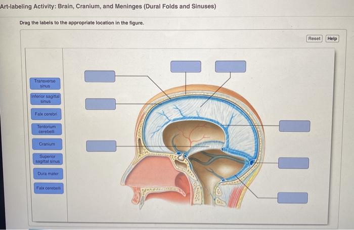
Solved Art-labeling Activity: Brain, Cranium, and Meninges ...

lab 7 (exercise 14) Flashcards | Quizlet
Associate Degree Nursing Physiology Review

9 BRAIN ideas | brain, dura mater, brain anatomy

The Spinal Cord Labeled Diagram Stock Photo, Picture And ...
0 Response to "38 drag the labels onto the diagram of the cns meninges."
Post a Comment