39 gel electrophoresis labeled diagram
Page Not Found Ap biology gel electrophoresis lab answers A diagram of the gel-banding pattern within the capillary ... A diagram of the gel-banding pattern within the capillary is to be drawn. Concept introduction: A laboratory technique that is used for the separation of charged molecules such as proteins, DNA and RNA on the basis of their size is known as gel electrophoresis. This technique is useful to distinguish DNA fragments of various lengths.
Allele-specific oligonucleotide - Wikipedia An allele-specific oligonucleotide (ASO) is a short piece of synthetic DNA complementary to the sequence of a variable target DNA. It acts as a probe for the presence of the target in a Southern blot assay or, more commonly, in the simpler Dot blot assay. It is a common tool used in genetic testing, forensics, and Molecular Biology research.. An ASO is typically an oligonucleotide of …
Gel electrophoresis labeled diagram
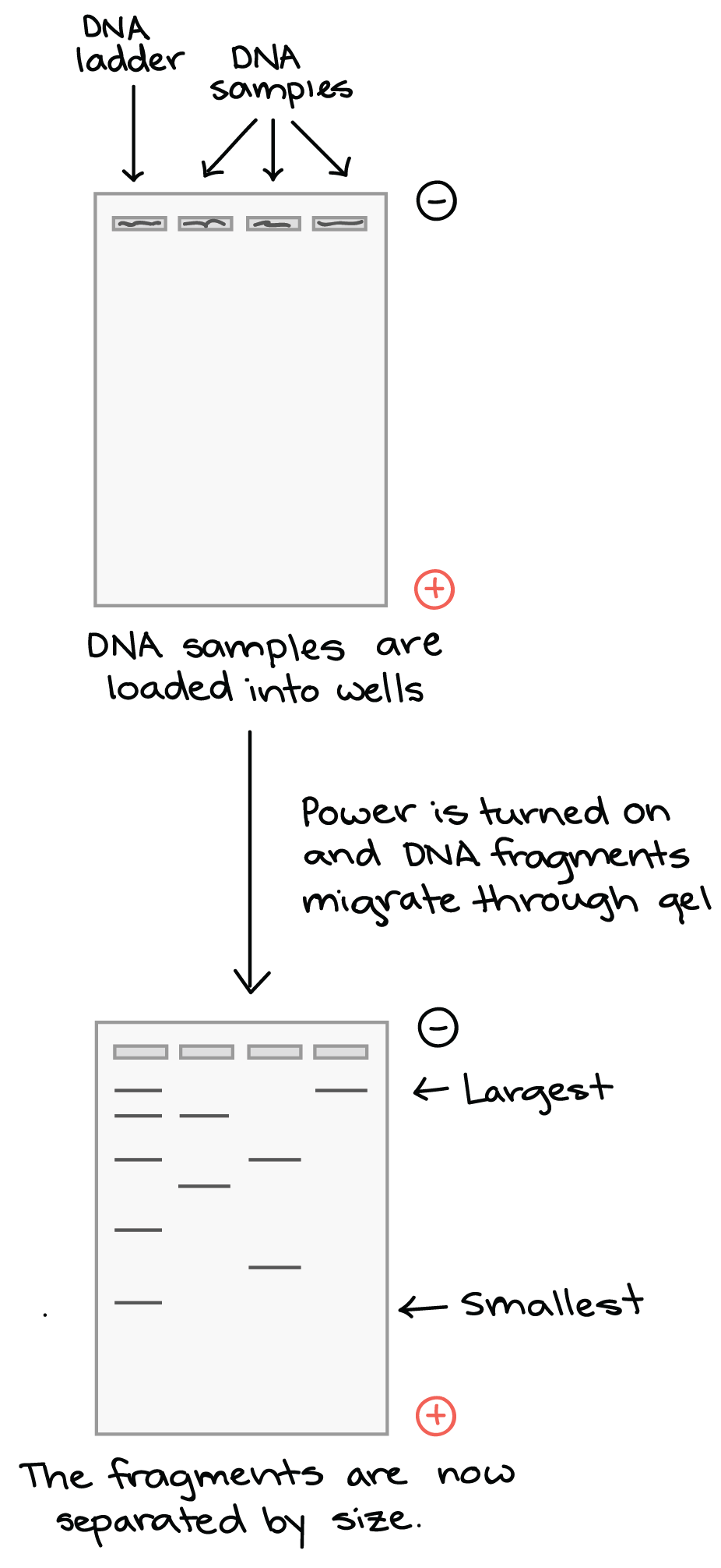
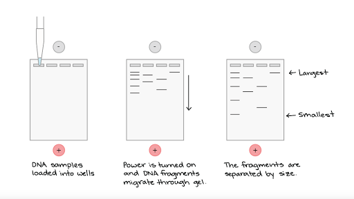
Techniques used in Molecular Biology (iii) Gel electrophoresis: Gel electrophoresis is one of the principal tools of molecular biology. The basic principle is that DNA, RNA, and proteins can all be separated by means of an electric field. In agarose gel electrophoresis, DNA and RNA can be separated on the basis of size by running the DNA through an agarose gel. PDF Lab 4: Gel Electrophoresis - Vanderbilt University Gel electrophoresis is a method of separating DNA fragments by movement through a Jello-like substance called agarose. Derived from a seaweed polysaccharide, agarose gels form small pores ... terms are labeled on the gel, and the loading key is labeled according to each lane. 1000bp 500bp 2000bp 250bp 100bp Lane 4 Lane Sample 1 DNA Ladder 2 ... Frog Dissection - Carolina.com Cut through the skin, following the pattern shown in the diagram below. Follow the same pattern to cut through the muscle and reveal the internal organs. Find the large brownish structure in the center of the body cavity, the liver. This is the largest internal organ that consists of 3 lobes. Lift the lobes of the liver and locate the gallbladder.
Gel electrophoresis labeled diagram. Mini Lesson 2 Gel Electrophoresis Answer the | Chegg.com Transcribed image text: Mini Lesson 2 Gel Electrophoresis Answer the following questions based upon the diagram of the gel seen below Lane 1: Standard Base pair ladder DNA-Hindill Digest Uncut = 48,502 bp 23,130 1 9,416 → Nm 6,557 → 4,361 →4 2,3225 2,027 6 7 564 17 125 →8 1. Label the positive and negative electrode side of the gel. 2. Draw an arrow on the gel in the direction the DNA ... PDF Gel Electrophoresis (Revised 3/8/2005) - Weebly or optical gel reading, it employed a laser to detect the fluorescently labeled nucleotides. The sequencer incorporated a computer program that builds a simulated gel image of colored DNA bands as they pass a scanning laser during electrophoresis. The final output took the form of an electropherogram, showing AP BIO REVIEW (unit 6) Flashcards - Quizlet Scientists conducted a transformation experiment using E. coli bacteria and the pTru plasmid. Samples of the pTru plasmid (lane A) and the chromosomal DNA from two different E. coli strains that the scientists attempted to transform (lane B and lane C) were compared using gel electrophoresis. The results are shown in Figure 1. Gel Electrophoresis - an overview | ScienceDirect Topics Gel Electrophoresis with Laser Ablation Applied to Cadmium Speciation in Proteins. Gel electrophoresis is a well-known separation technique for complex media such as proteins. However, classical modes of detection (including dye staining, immunoreaction with antisera, and autoradiography) do not allow the detection of metal-protein complexes.
43 gel electrophoresis labeled diagram Gel electrophoresis labeled diagram › biochemistryTop 12 Types of Chromatographic Techniques | Biochemistry Either technique should not be confused with gel electrophoresis, where an electric field is used to "pull" or "push" molecules through the gel depending on their electrical charges. SEC is a widely used technique for the ... Agarose gel electrophoresis of labeled DNA in which the ... Download scientific diagram | Agarose gel electrophoresis of labeled DNA in which the same gel is displayed before staining (Unstained) and after ethidium bromide staining (EtBr Stained). Lanes C ... Module 1.2: Agarose Gel Electrophoresis | Labs ... Diagram of agarose gel setup, for agarose gel electrophoresis. (Figure by MIT OpenCourseWare.) Today you will separate DNA fragments using an agarose matrix. Agarose is a polymer that comes from seaweed and if you've ever made Jell-O™, then you already have all the skills for pouring an agarose gel. Gel Electrophoresis Diagram Quiz - PurposeGames.com About this Quiz. This is an online quiz called Gel Electrophoresis Diagram. There is a printable worksheet available for download here so you can take the quiz with pen and paper.
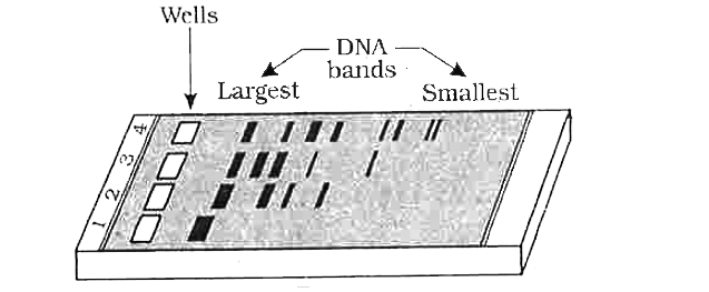
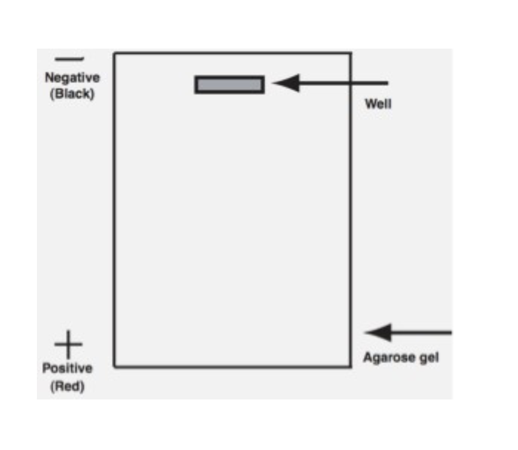
A complete guide for analysing and interpreting gel ... A Complete Guide for Analysing and Interpreting Gel Electrophoresis Results. Agarose gel electrophoresis is an important technique in molecular genetics for a long. DNA bands can only be visualized using agarose gel electrophoresis. In genomic research, analyzing and interpreting the agarose gel electrophoresis results are very crucial. Biology 101: Gel Electrophoresis - Moosmosis The gel is a porous matrix like a sponge and separates the DNA based on two main things: 1) size and 2) charge. Gel Electrophoresis: Separation By Size. As DNA is moved through the gel, smaller sized fragments move through faster than larger sized fragments. For example, a 100 base pair fragment will move through the gel faster than a 500 bp ... Solved tduring electrophoresis. On the gel diagram below ... Transcribed image text: tduring electrophoresis. On the gel diagram below, show how you believe these fragments will sort out Label each fragment with its correct number of base pairs. 7. 2,322 4,361 9,416 2.027 23,130 6,557 Well -Agarose gel Fountainhead Pres PDF Gel Electrophoresis: How Does It Work - Purdue University a. After you find out what dyes you are using, draw a picture of the gel and the wells. Label which dyes you will put in each well. b. When you load a gel, it is very important that you do not damage the gel in any way. You must be very careful not to "jab" the gel with the end of your pipet. Ideally, you shouldn't even touch the gel with the ...
Electrophoresis
BAM Chapter 17: Clostridium botulinum | FDA Prepare a 1.2-1.5 % agarose gel in 0.5 × TBE containing 0.5 µg ethidium bromide/ml agarose. Agarose may be melted in 0.5 × TBE using a microwave. Cast gel and allow to solidify.

InDesign Labeling / Annotating PCR Gel Pictures - Advanced Tutorial (Part 12)
PDF GEL ELECTROPHORESIS OF DYES - APS Home the solidified gel. Remove the tape from the ends of the gel tray. 6. Place gel into electrophoresis unit. Add 150 ml 1X TBE buffer to completely fill the box and to cover the top gel surface with about 2 mm of buffer. NOTE: At this point the gel box can be covered and left until the next day if necessary 7.

Agarose Gel Electrophoresis: Results Analysis Video
3 Ways to Read Gel Electrophoresis Bands - wikiHow Gel electrophoresis is a type of biotechnology that separates molecules based on their size to interpret an organism's DNA. An enzyme is used to separate a strand of DNA from a source and the DNA is suspended in a dye. Then, the dye is applied to a negatively-charged gel on one side of a sheet.

Agarose Gel Electrophoresis - an overview | ScienceDirect Topics
Chapter 7 Flashcards | Quizlet Label the diagram with the names of the three components of a nucleotide. Left: phosphate group Bottom Right: deoxyribose ... Why does DNA travel toward the positive electrode during gel electrophoresis? ☑ DNA is negatively charged, and opposite charges attract. ☐ DNA is neutral, but it associates with negatively charged RNA. ...

Agarose gel electrophoresis of DNA - Cleaver Scientific
MCB: Exam 2 Sapling Questions Flashcards - Quizlet The first lane in the gel electrophoresis diagram is a control lane, where no DNA‑binding protein was added. Lanes 2-4 are experimental lanes where one DNA‑binding protein was added to the DNA of interest prior to DNase I treatment. Identify whether each region of the DNA indicated on the gel is bound by a DNA‑binding protein.
Gel Electrophoresis - Definition, Purpose and Steps | Biology ...
Agarose Gel Electrophoresis for the Separation of DNA ... Agarose gel electrophoresis is the most effective way of separating DNA fragments of varying sizes ranging from 100 bp to 25 kb 1.Agarose is isolated from the seaweed genera Gelidium and Gracilaria, and consists of repeated agarobiose (L- and D-galactose) subunits 2.During gelation, agarose polymers associate non-covalently and form a network of bundles whose pore sizes determine a gel's ...
![SDS-gel electrophoresis of [ 35 S]methionine and [ 35 S ...](https://www.researchgate.net/profile/Uddhav-Kelavkar/publication/12133784/figure/fig3/AS:341752268509188@1458491498739/SDS-gel-electrophoresis-of-35-Smethionine-and-35-Scystine-labeled-proteins-isolated.png)
SDS-gel electrophoresis of [ 35 S]methionine and [ 35 S ...
PDF AP BIOLOGY 2007 SCORING GUIDELINES - College Board (a) Using the circle provided, construct a labeled diagram of the restriction map of the plasmid. Explain how you developed your map. Construct a labeled map and explain (3 points maximum) H H E E 40 20 30 10 H E 40 20 30 10 E H E = EcoRI Restriction Point H = HaeIII Restriction Point
PLOS ONE: Investigations on therapeutic glucocerebrosidases ...
Two-dimensional gel electrophoresis of L-[35S]methionine ... Download scientific diagram | Two-dimensional gel electrophoresis of L-[35S]methionine-labeled proteins in sulfur-free medium. MTD2-B (bcyl/bycl pSY2-1) (a), MTD2 from publication: The adenylate ...

Draw a neat labelled diagram of a typical agarose gel ...
The results of gel electrophoresis are shown below with ... Answer this q The results of gel electrophoresis are shown below, with four different strands of DNA labeled.Which strand of DNA is the shortest? uestion…
Bioanalytical chemistry 4. Gel Electrophoresis
The necessity of and strategies for improving confidence ... The western blot. The western blot, also sometimes referred to as the immunoblot, involves separating native or denatured proteins by gel electrophoresis, transferring these separated proteins to a protein binding membrane and subsequent detection of a target protein by an antibody specific to the target protein (Figure 1).Although western blotting is mainly carried out …

Gel electrophoresis of nucleic acids - Wikipedia
3 Main Types of Chromatography Techniques (With Diagram) Electrophoresis: Electrophoresis was invented by Tiselius in 1937. It is based on the principle that the proteins in a gel migrate in an electric field till they reach their isoelectric point. Isoelectric point is the pH of the medium at which their net charge is zero. Electrophoresis through a gel separates DNA, RNA and protein molecules.
Draw a neat labelled diagram of a typical agarose gel ...
Gel electrophoresis (article) - Khan Academy Gel electrophoresis. AP.BIO: IST‑1 (EU) , IST‑1.P (LO) , IST‑1.P.1 (EK) A technique used to separate DNA fragments and other macromolecules by size and charge. Google Classroom Facebook Twitter.

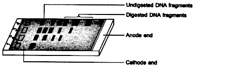
Figure legends Figure 1: Agarose gel electrophoresis (2 ...
How to Interpret DNA Gel Electrophoresis Results | GoldBio The next step is to identify those bands to figure out which one to cut. Gel Electrophoresis. Lane 1: DNA Ladder. Lane 2: Undigested plasmid A. Lane 3: Completely digested plasmid A. Lane 4: Digested PCR product (or DNA Fragment). Lane 5: PCR Product (with a faint primer dimer band). Lane 6: Genomic DNA.

1% agarose gel electrophoresis showing the amplified RT-PCR ...
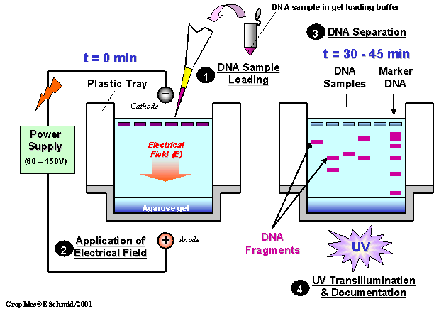
Electrophoresis 1 and 2 Lab (1).pdf - Lab 10 ... 82 BIOLOGY 1107 5. Running your gel a. Make sure the power supply is off before connecting it to the gel box. b. Carefully place the lid on the electrophoresis chamber and match up the + and - electrodes accordingly at the power supply. c. Check the orientation of the gel with how you labeled your diagram in Figure 1.

Confirmation of QD-labeling of pDNA by agarose gel ...
Electrophoresis (With Diagram) - Biology Discussion Electrophoresis (With Diagram) The term electrophoresis describes the migration of a charged particle under the influence of an electrical field. Many important biomolecules — such as peptide, proteins nucleotides and nucleic acids — possess ionisable groups and, therefore, at any given pH, exist in solution as electrically charged species ...

Electrophoresis and Blotting of DNA - Tamber - - Major ...
Gel Electrophoresis Diagram | Quizlet Start studying Gel Electrophoresis. Learn vocabulary, terms, and more with flashcards, games, and other study tools.
12.2: Visualizing and Characterizing DNA - Biology LibreTexts
On the gel diagram at the right show how you believe these ... Place your gel on a light background and record your results by making a diagram as follows. Place a clear sheet of plastic sheet or acetate over the gel. With a permanent marker, trace the wells and band patterns onto the plastic sheet to make a replica picture of your gel. Remove the plastic sheet for later analysis.

Answer in 3 to 5 sentences:Draw a neat labelled diagram of a ...
Protein Separation by Capillary Gel Electrophoresis: A ... Jan 04, 2012 · 1. Introduction. Sodium dodecyl sulfate-polyacrylamide gel electrophoresis (SDS-PAGE, see Table I for a list of acronyms used in this paper) has been used for size-based separations of proteins for over four decades [1, 2], and it is still the workhorse for protein separations and analyses in most biological research laboratories.The basic procedures of this …

SOLVED:Complete this rule for the movement of DNA fragments ...
Answered: On the gel diagram below, show how you… | bartleby Science Biology Q&A Library On the gel diagram below, show how you believe these fragments will sort out during electrophoresis. Label each fragment with its correct number of base pairs. Label each fragment with its correct number of base pairs.

Two-Dimensional Gel Electrophoresis - an overview ...
Answer in 3 to 5 sentences:Draw a neat labelled diagram of ... In agarose gel electrophoresis, agarose is used as a matrix. The sample is added in the slot and current is applied to it. The smaller molecules move faster and the larger molecules are retarded. In this method, separation is based on charge and size of the molecules. DNA, RNA and proteins can be separated using this technique.

Draw a neat labelled diagram of a typical agarose gel ...
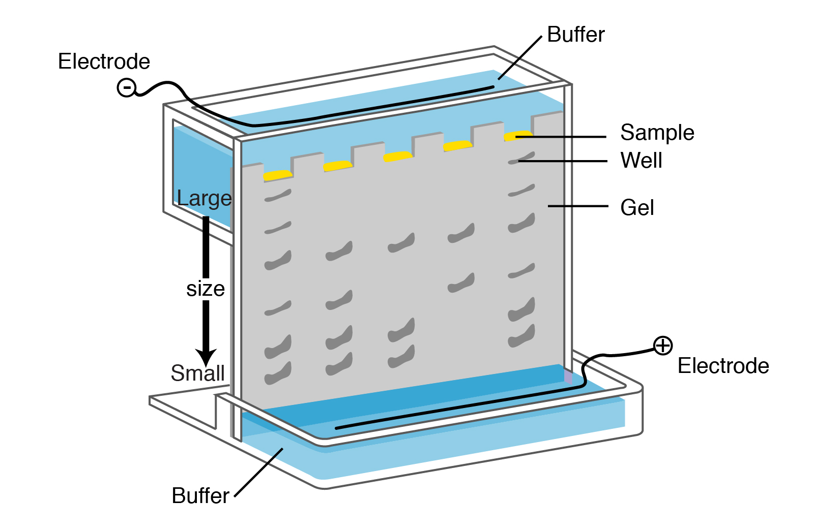
Recombinant DNA Technology (With Diagram) Gel electrophoresis employs a buffer system, a medium which is a gel and a source of direct current (Fig. 6). Samples having DNA fragments are applied on the gel and current is passed through the system for an appropriate time. Different DNA fragments move up to different distances on the gel depending on their charge to mass ratio. The heavier ...
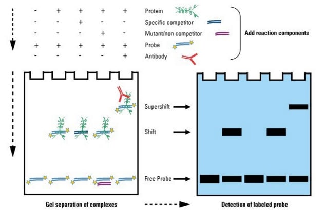
Gel Shift Assays (EMSA) | Thermo Fisher Scientific - US
Frog Dissection - Carolina.com Cut through the skin, following the pattern shown in the diagram below. Follow the same pattern to cut through the muscle and reveal the internal organs. Find the large brownish structure in the center of the body cavity, the liver. This is the largest internal organ that consists of 3 lobes. Lift the lobes of the liver and locate the gallbladder.

Gel electrophoresis (video) | Khan Academy
PDF Lab 4: Gel Electrophoresis - Vanderbilt University Gel electrophoresis is a method of separating DNA fragments by movement through a Jello-like substance called agarose. Derived from a seaweed polysaccharide, agarose gels form small pores ... terms are labeled on the gel, and the loading key is labeled according to each lane. 1000bp 500bp 2000bp 250bp 100bp Lane 4 Lane Sample 1 DNA Ladder 2 ...

cont.) GEL ELECTROPHORESIS (basic concept) - ppt video online ...
Techniques used in Molecular Biology (iii) Gel electrophoresis: Gel electrophoresis is one of the principal tools of molecular biology. The basic principle is that DNA, RNA, and proteins can all be separated by means of an electric field. In agarose gel electrophoresis, DNA and RNA can be separated on the basis of size by running the DNA through an agarose gel.
Gel Electrophoresis: Basics & Steps | SchoolWorkHelper

Activity 2 - Gel Electrophoresis of Dyes

Given below is the diagram representing the observations made ...

Module 1.2: Agarose Gel Electrophoresis | Labs | Laboratory ...
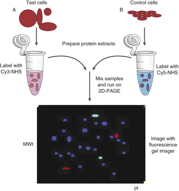
Two-dimensional difference gel electrophoresis | Nature Protocols

Evaluation of PCR-labeled probes by agarose gel ...
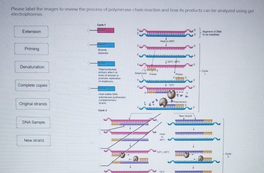
Solved Please label the images to review the process of ...

Biology: Chapter 21 - DNA profiling (diagram) Diagram | Quizlet
Use the gel diagram to indicate how you believe the | Chegg.com
Gel electrophoresis (article) | Khan Academy

Gel Electrophoresis Diagram | Quizlet
Gel electrophoresis (article) | Khan Academy

Gel electrophoresis of 13Hjproline-labelled, pepsin-digested ...
Draw a diagram of a typical agarose gel electrophoresis ...

Evaluation of PCR-labeled probes by agarose gel ...
0 Response to "39 gel electrophoresis labeled diagram"
Post a Comment