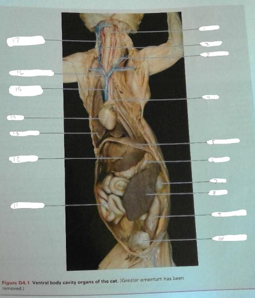
39 cat veins and arteries diagram
Axillary vein (Vena axillaris) The axillary vein is a deep vein of the upper limb that is formed by the union of the brachial and basilic veins.It starts at the lower border of the teres major muscle and ascends medially through the axilla towards the 1st rib, where it is continued by the subclavian vein.. Along its course, the axillary vein lies anteromedial to the axillary artery, partially ... Cat Arteries Diagram | Quizlet Cat Arteries STUDY Learn Write Test PLAY Match + − Created by elosasloth PLUS Terms in this set (17) aortic arch ... brachiocephalic artery ... left common carotid artery ... right common carotid artery ... right subclavian artery ... left subclavian artery ... axillary artery ... brachial artery ... celiac trunk ...
just a bunch of different cat artery and vein diagrams to practice on! I tried to compile as many games as I could find so this should be a compilation of most (if not all) cat vein and arteries games. choose your favorite. Play All Use in Tournament Branch. Major Veins and Arteries of Cat (Upper Thorax) by xglued 19,741 plays 25p Image Quiz. Cat Arteries and Veins by Drewlojo 7,451 plays 43p ...
Cat veins and arteries diagram
Neck spaces. The content of the neck is grouped into 4 neck spaces, called the compartments.. Vertebral compartment: contains cervical vertebrae and postural muscles.; Visceral compartment: contains glands (thyroid, parathyroid, and thymus), the larynx, pharynx and trachea.; Two vascular compartments: contain the common carotid artery, internal jugular vein and the vagus nerve, on each side of ... Anatomy of the arteries and bones of the lower limb based on 3D pictures and angiogram (angiography). This part of the interactive atlas of anatomy of the human body is about the arterial vasculature of the pelvic girdle, pelvis, thigh, knee, leg and foot and the study of bones and joints. It includes a 3D reconstruction of bones and arteries ... The hyoid bone is a small horseshoe-shaped bone located in the front of your neck. It sits between the chin and the thyroid cartilage and is instrumental in the function of swallowing and tongue movements. 1 . The little talked about hyoid bone is a unique part of the human skeleton for a number of reasons. First, it's mobile.
Cat veins and arteries diagram. Pulmonary vein carries the blood from the lungs to the left atrium. The artery is the vessel which carries the blood from the heart towards the body. Artery contains pure blood i.e. oxygen mixed blood. But Pulmonary arteries are the exception which always carries the impure blood or deoxygenated blood. Diagrams of Feline Arterial and Venous Systems Arterial System Diagram. Venous System Diagram. Posted by Unknown at 9:10 PM. Labels: Anatomy and physiology, arteries, cat, feline, veins. 2 comments: PureandBeautiful.com said... Thank you! I appreciate your posting the cat pics. I have looked all over the web for blank pics and yours are the only ones. THANKX!!!! November 4, 2011 at 5:06 PM ... Google Health wants to help everyone live more life every day. Learn more about what Google Health is and how we're aiding in healthcare advancements. Pseudofracture of the apophysis is a partially ossified apophyseal plate that appears on an X-ray as if it was a fracture. This is a normal variant of the calcaneus and does not require treatment. Congenital tarsal coalition is a connection between tarsals, usually the calcaneus and talus, that prevent the tarsals from articulating properly. The coalition can be from ossification (bone fusion ...
Blood is transported in arteries, veins and capillaries. Blood is pumped from the heart in the arteries. It is returned to the heart in the veins. The capillaries connect the two types of blood ... A pulmonary embolism is a blood clot that becomes lodged in the pulmonary arteries. The majority of emboli arise because of deep vein thrombosis in the legs. Pulmonary emboli may be investigated using a ventilation/perfusion scan, a CT scan of the arteries of the lung, or blood tests such as the D-dimer. The profile of the rear quarters of a healthy cat should give the impression of strength and support. The body profile will taper down slightly toward the tail end, while remaining well-muscled, particularly around the haunches. A slight belly pouch is normal, although it is more prominent in heavier cats, or in obese cats who have lost weight. Human heart is basically a biological pump that circulates blood in the human body. The heart is the size of a human fist and is located between the lungs in the thoracic cavity, slightly towards the left of the sternum (breastbone). It forms the cardiovascular system together with blood vessels, veins and capillaries.
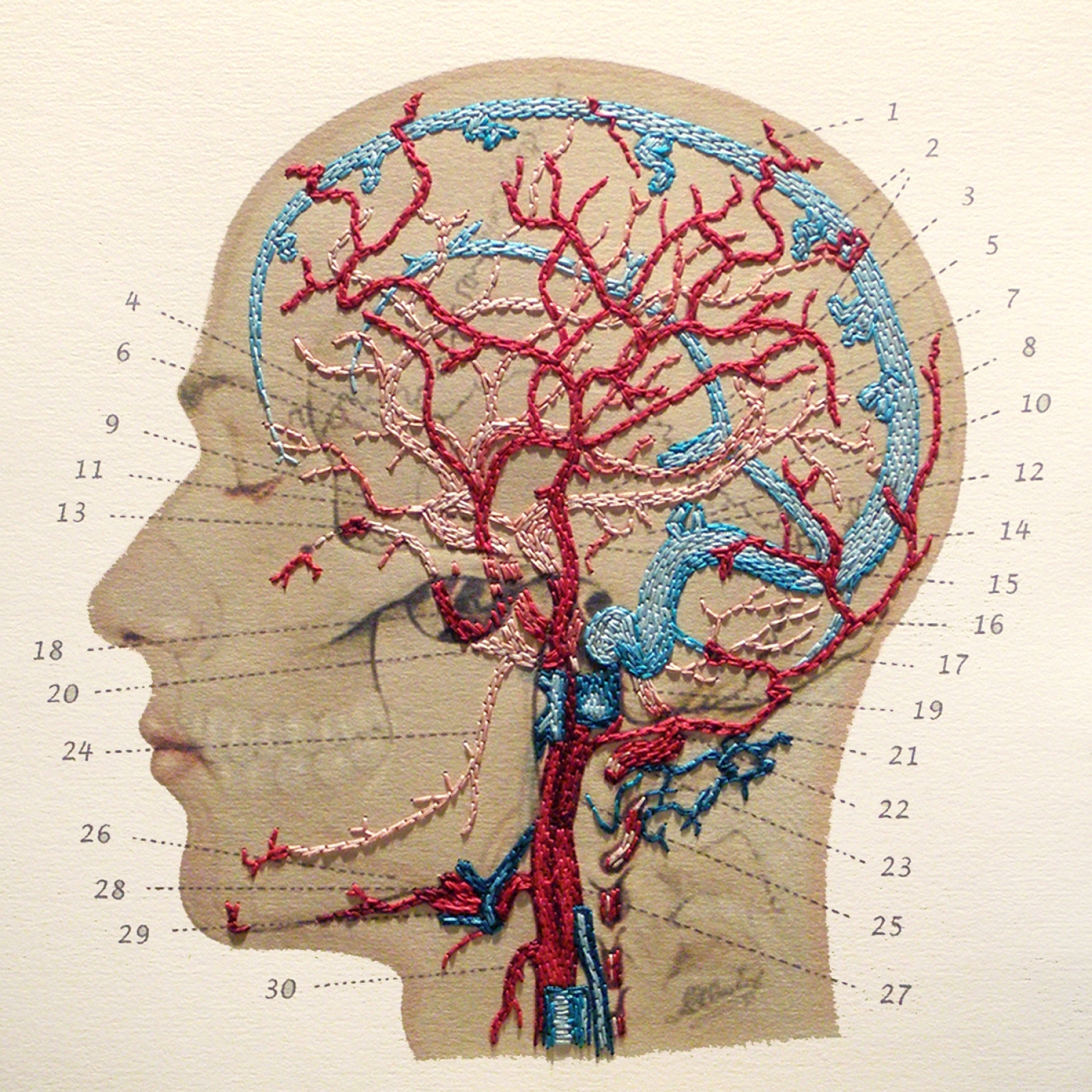
Magnetic resonance imaging (MRI) is a medical imaging technique used in radiology to form pictures of the anatomy and the physiological processes of the body. MRI scanners use strong magnetic fields, magnetic field gradients, and radio waves to generate images of the organs in the body. MRI does not involve X-rays or the use of ionizing radiation, which distinguishes it from CT and PET scans. Veins are darker and slightly wider than corresponding arteries. The optic disc is at right, and the macula lutea is near the centre. The retina is stratified into distinct layers, each containing specific cell types or cellular compartments [36] that have metabolisms with different nutritional requirements. [37] DIGETIVE SYSTEM - CAT & MODELS, HEART, ARTERIES, EKG, BLOOD etc. (These videos are meant to help visually review some content - please follow book and instructions from your professor as well) CAT ARTERIES by GARY MOO YOUNG (PLAYLIST) CAT VEINS by GARY MOO YOUNG (PLAYLIST) BSC2086L AP2 lab FINAL LAB EXAM E-BOOK Each posterior cerebral artery sends a small posterior communicating artery to join with the internal carotid arteries. Blood drainage. Cerebral veins drain deoxygenated blood from the brain. The brain has two main networks of veins: an exterior or superficial network, on the surface of the cerebrum that has three branches, and an interior network.
Arteries are the blood vessels that carry blood away from the heart, where it branches into even smaller vessels. Finally, the smallest arteries, called arterioles are further branched into small capillaries, where the exchange of all the nutrients, gases and other waste molecules are carried out.. Veins are the blood vessels present throughout the body.
The urinary bladder and urethra are pelvic urinary organs whose respective functions are to store and expel urine outside of the body in the act of micturition (urination). As is the case with most of the pelvic viscera, there are differences between male and female anatomy of the urinary bladder and urethra. In our entire urinary system series, the urinary bladder and urethra represent the ...

Image from page 380 of "Anatomical technology as applied to the domestic cat; an introduction to human, veterinary, and comparative anatomy" (1882)
Many dogs slowly develop degenerative thickening and progressive deformity of one or more heart valves as they age. In time, these changes cause the valve, most commonly the mitral valve, to leak. While it can lead to heart failure, many dogs with the condition never show signs of the disease outside of a loud heart
Calcification occurs when deposits of calcium form in the body. There are many types, each with their own causes, symptoms, and treatments. Find out more.
Double Circulation Diagram. Fig: Double Circulation. Learn 12th CBSE Exam Concepts. Components Involved in Double Circulation. 1. Heart- It is four-chambered in humans, i.e. right and left atrium & right and left ventricle. 2. Blood vessels- Arteries, veins and capillaries come under blood vessels. Arteries carry oxygenated blood; veins ...
From the tissues that derive from the embryonic ectoderm and endoderm, we turn now to those derived from mesoderm. This middle layer of cells, sandwiched between ectoderm and endoderm, grows and diversifies to provide a wide range of supportive functions. It gives rise to the body's connective tissues, blood cells, and blood vessels, as well as muscle, kidney, and many other …
10 terms meghantsmythPLUS Cat - Arteries and Veins 4 STUDY transverse jugular vein ... left external jugular vein ... left internal jugular vein ... left brachiocephalic vein ... right brachiocephalic vein ... internal mammary vein ... superior vena cava ... left subclavian vein ... left axillary vein ... left brachial vein ...
Home Sheep Heart Labeled Diagram Sheep Heart Labeled Diagram. NoName Dec 30, 2021 Dec 30, 2021
Cardiology is that branch of medicine which deals with the diagnosis and treatment of heart diseases. Cardiologists investigate patients with suspected heart disease by taking a very careful, extensive history of the patient's condition, and performing a complete physical examination.
Capillaries are very tiny blood vessels that form a connection between arteries and veins. The capillary walls facilitate the transfer of oxygen, nutrients and wastes in and out of your body. 3. The Lungs. Your lungs aren't technically a part of circulatory system organs, but they really help make it possible for your heart to function ...
On the left is an anatomy diagram of the internal organs of a female elephant. Click on the image for a larger look at it. The larger image will open in a new window, use the close button when finished. Below you can see some distinct differences between the African elephant and the Asian elephants body structures.
Arterial supply: approximately 10 arterial branches from anastomotic arcade of ovarian artery (branch of aorta) and ovarian branch of uterine artery penetrate hilus into medulla and cortex Venous drainage: left ovarian vein drains to left renal vein, right ovarian vein drains to inferior vena cava

The Return of Odysseus (Homage to Pinturicchio and Benin) (1977) // Romare Howard Bearden American, 1911-1988
On the image below the arteries are colored pink. The veins visible at the top of the heart include the superior vena cava, the brachiocephalic veins (2) and the jugular. This diagram was useful for understanding the layout of these vessels.
Activity 2.1 Directions: Complete the diagram below by choosing the correct words from the box below. Circulatory System N 1 HEART WBC 3 4 5 VEINS CO …
Cat Dissection Muscles. 31 terms. hg00829p. Muscles of the Lower Extremity. 41 terms. robswatski TEACHER. cat veins and arteries. 39 terms. juliadesantis1. Sets with similar terms. Cat Muscles (Images) 53 terms. meliciementor. Identify the Gender - 3rd declensions. 87 terms. Yonatan_Gut. RAD105-1 Facial Pictures.
This is an online quiz called Cat Arteries and Veins. There is a printable worksheet available for download here so you can take the quiz with pen and paper. Your Skills & Rank. Total Points. 0. Get started! Today's Rank--0. Today 's Points. One of us! Game Points. 43. You need to get 100% to score the 43 points available. Actions. Add to favorites 8 favs. Add to Playlist 5 playlists. Add to ...
Like the arteries and the veins, it lies superficially located between the tunica albuginea and the clitoral fascia, and therefore some procedures (e.g. vulvoplasty) may risk injury to this nerve and affect clitoral sensation and sexual function. Function. During sexual arousal, the clitoris, along with the entire female genitalia, fills with ...
With a program that lets you explore your scientific interests, gain real-world experience, and make valuable connections with faculty experts, you’ll turn your passion for science into the start of a fulfilling career. Internationally recognized. Internally driven. In seven different buildings ...
The Demon core is a 6.2 kg Plutonium-Gallium sphere with a diameter of 8.9 cm (smaller than a basketball). The Plutonium-239 isotope used in the core is a manmade element produced in nuclear reactors. It is highly unstable, radioactive, and decays by emitting alpha particles, which is damaging to our tissues. Hence, the core has a nickel coating, which blocks the short-range alpha particles ...
Oct 01, 2017 · Circulatory System. Cat Upper Neck Arteries Veins Diagram Quizlet. Cat Veins Purposegames. Https Massasoit Instructure Com Courses 902777 Files 29763690 Download Wrap 1. Anatomy Lab Practicum 3 Cat Arteries And Veins Diagram Quizlet. Diagrams Cat Veins And Arteries Diagram. Untitled Document. Labeled Cat Veins Diagram.
Arteries - Arteries are vessels that carry blood away from the heart. Veins - Veins bring blood back to the heart. Capillaries - Capillaries are fine vessels that connect the arteries and veins. Circulatory System: Lymphatic system. The lymphatic system is a network of tissues and organs that helps the body in getting rid of toxins, waste and other unwanted materials.
Cat arteries (diagram) 15 terms. here4um8 PLUS. ... 2 terms. here4um8 PLUS. Hepatic portal veins (diagram) 5 terms. here4um8 PLUS. Caudal veins pt 4. 5 terms ...
As with the mouse data, the analysis was restricted to non-cycling arteries, capillaries, and veins in order to specifically compare cell states and trajectories in coronary vessels. Similar to mouse, the data contained one vein and two capillary clusters, but in contrast to mouse, there was an additional arterial cluster for a total of three ...
There are two types of them: arteries and veins. Arteries: These types of blood vessels take oxygen-rich blood from the heart and transport to the capillaries. Arteries are quite tough on the outside but are smooth on the inside. There are three arteries of the heart, including pulmonary artery, aorta, and coronary arteries.
Significance of the Paradox. Theoretically, the earth must have frozen under a faint, young sun. However, the earth remained warm and toasty even when the sun was dim. This could be due to a higher concentration of greenhouse gases or due to the lower albedo of the early earth. 4.5 billion years ago, in the vast expanse of space, gas clouds ...
Cat dissection labeled arteries and veins. We remove the pericardium from the heart and carefully tease away the connective tissue to reveal the arteries that leave the heart. Superior mesenteric artery renal arteries external iliac arteries femoral vein femoral arteries internal iliac arteries common iliac veins abdominal aorta renal veins. Online quiz to learn major veins and arteries of cat ...
25.1.2018 · Arteries. Arteries are blood vessels that transport oxygenated blood from the heart to various parts of the body. They are thick, elastic and are divided into a small network of blood vessels called capillaries. The only exception to this is the pulmonary arteries, which carries deoxygenated blood to the lungs. Veins
Arteries and veins of the thoracic wall. The thoracic wall or chest wall is a musculoskeletal structure that has a vast vascular supply. Most of the arteries of the thoracic cavity arise directly from the thoracic aorta; while others arise from its branches.On the other hand, the veins of the thoracic wall eventually coalesce to drain into the vena caval system.

Image from page 263 of "The cat : an introduction to the study of backboned animals, especially mammals" (1881)
Insect morphology is the study and description of the physical form of insects.The terminology used to describe insects is similar to that used for other arthropods due to their shared evolutionary history. Three physical features separate insects from other arthropods: they have a body divided into three regions (called tagmata) (head, thorax, and abdomen), have three pairs …
The immune system is a network of biological processes that protects an organism from diseases.It detects and responds to a wide variety of pathogens, from viruses to parasitic worms, as well as cancer cells and objects such as wood splinters, distinguishing them from the organism's own healthy tissue.Many species have two major subsystems of the immune system.
Cats are scared of cucumbers because it's their natural reaction to anything that sneaks up on them without making any noise. Cats tend to be scared or wary of the unknown, The internet is a bizarre place. If you take the time, 5 hours or so, you'll get sucked into the powerful Charybdis-like whirlpool of internet content.
The hyoid bone is a small horseshoe-shaped bone located in the front of your neck. It sits between the chin and the thyroid cartilage and is instrumental in the function of swallowing and tongue movements. 1 . The little talked about hyoid bone is a unique part of the human skeleton for a number of reasons. First, it's mobile.
Anatomy of the arteries and bones of the lower limb based on 3D pictures and angiogram (angiography). This part of the interactive atlas of anatomy of the human body is about the arterial vasculature of the pelvic girdle, pelvis, thigh, knee, leg and foot and the study of bones and joints. It includes a 3D reconstruction of bones and arteries ...
Neck spaces. The content of the neck is grouped into 4 neck spaces, called the compartments.. Vertebral compartment: contains cervical vertebrae and postural muscles.; Visceral compartment: contains glands (thyroid, parathyroid, and thymus), the larynx, pharynx and trachea.; Two vascular compartments: contain the common carotid artery, internal jugular vein and the vagus nerve, on each side of ...








0 Response to "39 cat veins and arteries diagram"
Post a Comment