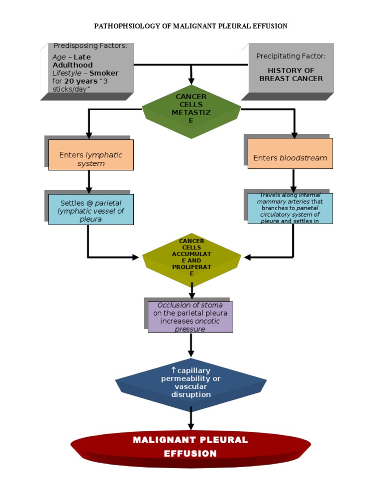
35 pleural effusion pathophysiology diagram
10.02.2017 · These diseases include pneumothorax, pleural effusion, or respiratory muscle weakness, such as can occur with botulism, tick paralysis, tetanus, strychnine poisoning, or severe white muscle disease. Ventilation-perfusion mismatches occur when the distribution of blood flow in the lungs does not match the distribution of alveolar ventilation, with the result … Pleural effusion, sometimes referred to as "water on the lungs," is the build-up of excess fluid between the layers of the pleura outside the lungs. The pleura are thin membranes that line the lungs and the inside of the chest cavity and act to lubricate and facilitate breathing. Normally, a small amount of fluid is present in the pleura.
Understanding pleural effusion. Nursing2004: August 2004 - Volume 34 - Issue 8 - p 64. Buy; Abstract In Brief. Find out how to recognize and manage excess fluid in the pleural space. Sort out signs and symptoms, possible underlying causes, and treatment options for this potential emergency.

Pleural effusion pathophysiology diagram
A malignant pleural effusion (MPE) is the build up of fluid and cancer cells that collects between the chest wall and the lung. This can cause you to feel short of breath and/or have chest discomfort. It is a fairly common complication in a number of different cancers. pathophysiology pleural effusion diagram. buy aspira pleural drainage. biapical nodular pleural scarring. mild basilar pleural thickening trauma. left apical pleural thickening. pictures of children postures. mild apical pleural. right pleural friction rub. mild bilateral apical pleural parenchymal scarring. edema in pleural cavity nodules A pleural effusion is collection of fluid abnormally present in the pleural space, usually resulting from excess fluid production and/or decreased lymphatic absorption. [] It is the most common manifestation of pleural disease, and its etiologies range in spectrum from cardiopulmonary disorders and/or systemic inflammatory conditions to malignancy.
Pleural effusion pathophysiology diagram. Chapter 128 Pleura & Pleural Space PLEURAL EFFUSION osms.it/pleural-effusion PATHOLOGY & CAUSES Excess fluid accumulates in pleural space Lung expansion limited → impaired ventilation Origin Hydrothorax (serous fluid), hemothorax (blood), urinothorax (urine), chylothorax/ lymphatic effusion (chyle), pyothorax (pus, AKA empyema) Pathophysiology Transudative pleural effusion Pressure driven ... Pathophysiology of Disease - An Introduction to Clinical Medicine, 7th Ed Objective: To review the pathophysiology and management of pleural effusions, including available agents for pleural sclerosis. Data sources: A MEDLINE search (1966 to present) was performed that included clinical studies in the English language involving the pathophysiology and management of pleural effusions; references used in those articles were screened for additional published information. Nov 30, 2021 · Pleural effusion pathophysiology diagram. 15 Oct 2021 — A pleural effusion is an abnormal collection of fluid in the pleural space resulting from excess fluid production or decreased absorption or ... Pneumonia is an inflammatory condition of the lung primarily affecting the small air sacs known as alveoli.
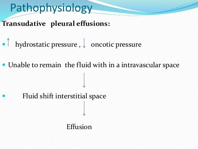
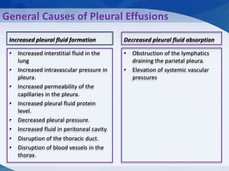
5 Pathophysiology (contd.) Normal pleural fluid 0.1 to 0.2 ml/kg Clear Low protein (1.0 to 1.5 g/dl) < 1500 nucleated cells / L 61% to 77% monocytes-macrophages 9 to 30% mesothelial cells 7% to 11% lymphocytes 2% neutrophils 0% eosinophils pH > 7.60 Pathophysiology (contd.) Mechanism of abnormal pleural fluid formation Increasedhydrostaticpressure(CHF)Increased hydrostatic pressure (CHF) Sep 01, 2021 · Pathophysiology. Pleural effusions develop when changes in fluid and solute homeostasis occur, and the mechanism causing these changes determines whether it will be an exudative (high protein content) or transudative (low protein content) effusion. Two features of human parietal pleura explain its role in the formation and removal of pleural liquid and protein in the normal state: the proximity of the microvessels to the pleural surface and the presence of stomata situated between mesothelial cells. Pathophysiology. Pleural effusion results either from increased pleural fluid formation or decreased exit of fluid. Increased Pleural Fluid Formation. Increased hydrostatic pressure (e.g. seen in congestive heart failure) Decreased colloid osmotic pressure (e.g. cirrhosis and nephrotic syndrome)
Where do I get my information from: http://armandoh.org/resourceFacebook:https://www.facebook.com/ArmandoHasudunganSupport me: http://www.patreon.com/armando... Definition Pleural effusion is a collection of abnormal amount of fluid in the pleural space. 4. Classification Transudative effusions Exudative effusions. 5. Transudative effusions Transudative effusions also known as hydrothoraces , occur primarily in noninflammatory conditions; is an accumulation of low-protein, low cell count fluid. 6. • Pleural Effusion: • Inflammatory Pleural Effusions: Serous, serofibrinous, and fibrinous pleuritis. • Noninflammatory Pleural Effusions: Noninflammatory collections of serous fluid within the pleural cavities are called hydrothorax. • The presence of blood into the pleural cavity is known as hemothorax. It is usually a complication of a ruptured aortic aneurysm or vascular trauma ... Attribution Non-Commercial (BY-NC) Available Formats. Download as DOC, PDF, TXT or read online from Scribd. Flag for inappropriate content. Download now. Save Save Pathophysiology- Pleural Effusion For Later. 75% (4) 75% found this document useful (4 votes) 6K views 1 page.
Pleural effusion is an indicator of an underlying disease process that may be pulmonary or nonpulmonary in origin and may be acute or chronic. Although the etiologic spectrum of pleural effusion is extensive, most pleural effusions are caused by congestive heart failure, pneumonia, malignancy, or pulmonary embolism 5.
A pleural effusion is an excessive accumulation of fluid in the pleural space. It can pose a diagnostic dilemma to the treating physician because it may be related to disorders of the lung or pleura, or to a systemic disorder. Patients most commonly present with dyspnea, initially on exertion, predominantly dry cough, and pleuritic chest pain.
Understood. This brings up some very interesting questions about the etiology of pleural effusion. In theory, your diagram should show where the fluid/matter is coming from. Can't really do that if you are creating a single diagram.
20.09.2014 · Nursing Care Plan for Dysphagia : Impaired Swallowing - These days we want to discuss the article with the title health Nursing Care Plan for Dysphagia : Impaired Swallowing we hope you get what you're looking for. We are here trying to make the best possible to provide information on this blog. Nursing Care Plan for Dysphagia : Impaired Swallowing
pleural effusion, also called hydrothorax, accumulation of watery fluid in the pleural cavity, between the membrane lining the thoracic cage and the membrane covering the lung.There are many causes of pleural effusion, including pneumonia, tuberculosis, and the spread of a malignant tumour from a distant site to the pleural surface. Pleural effusion often develops as a result of chronic heart ...
Chart and Diagram Slides for PowerPoint - Beautifully designed chart and diagram s for PowerPoint with visually stunning graphics and animation effects. Our new CrystalGraphics Chart and Diagram Slides for PowerPoint is a collection of over 1000 impressively designed data-driven chart and editable diagram s guaranteed to impress any audience.

Trading crypto with Coinberry in Canada! Via: techdaily.ca | #crypto #bitcoin #stocks #blockchain #dogecoin
11.01.2017 · Pleural Effusion.—Pleural effusion is seen in approximately 25% of primary tuberculosis cases in adults, with the vast majority of such effusions being unilateral . Pleural effusion is less common in children and may only appear in 6%–11% of pediatric cases, with increasing prevalence with age (2,20).
Two features of human parietal pleura explain its role in the formation and removal of pleural liquid and protein in the normal state: the proximity of the microvessels to the pleural surface and the presence of stomata situated between mesothelial cells. For pleural fluid to accumulate in disease, there must be increased production from increased hydrostatic pressure, decreased oncotic or ...
Pleural effusions are very common, and physicians of all specialties encounter them.A pleural effusion represents the disruption of the normal mechanisms of formation and drainage of fluid from the pleural space.A rational diagnostic workup, emphasizing the most common
A pleural effusion is the presence of an abnormal amount of fluid in the pleural space (a potential space between the visceral and parietal pleura). Pleural effusions can be transudative (lower protein/LDH) or exudative (higher protein/LDH).
Pneumonia is an inflammatory condition of the lung primarily affecting the small air sacs known as alveoli. Symptoms typically include some combination of productive or dry cough, chest pain, fever, and difficulty breathing. The severity of the condition is variable. Pneumonia is usually caused by infection with viruses or bacteria, and less commonly by other microorganisms.
The presence of caseating granulomas hypothyroidism pleural effusion pathophysiology diagram acid-fast bacilli on histological examination of the pleural surface is diagnostic of TB pleuritis 9. The signs usually include dyspnea, tachypnea, depression, anorexia, and weight loss.
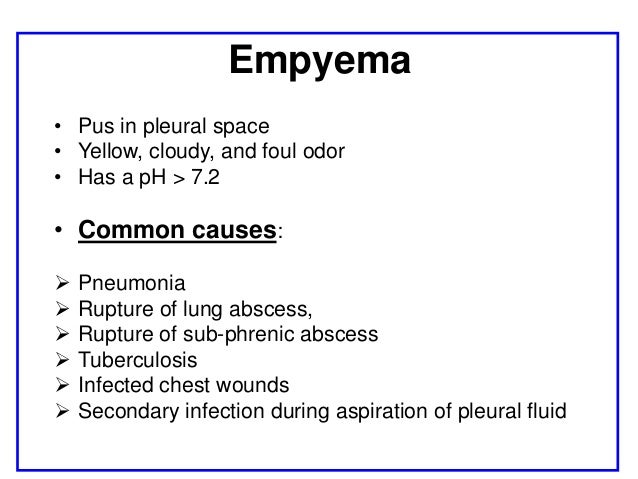
Pleural effusion. inflammation leads to exudation of fluid into pleural space. can compromise lung function. Empyema. purulent exudate in pleural space. necrosis/breakdown of visceral pleura and/or spread of infection into pleura. P. leural adhesions, lung fibrosis. Complications of pneumonia. Content Slide – Text w/ image. Logo: 1” high by ...
With large emboli; pleural friction rub, pleural effusion, fever, leukocytosis; Evaluation: Diagnosis can be made based on a patient's symptoms, medical history and a series of tests and scans. Clinical Decision Rules, such as the Well's Score, can guide diagnostics of suspected acute venous thromboembolism.
08.01.2014 · NCP for Hypoglycemia - Nursing Diagnosis and Interventions - These days we want to discuss the article with the title health NCP for Hypoglycemia - Nursing Diagnosis and Interventions we hope you get what you're looking for. We are here trying to make the best possible to provide information on this blog. NCP for Hypoglycemia - Nursing Diagnosis and …
Pleural effusion is suspected in patients with pleuritic pain, unexplained dyspnea, or suggestive signs. Diagnostic tests are indicated to document the presence of pleural fluid and to determine its cause (see figure Diagnosis of Pleural Effusion Diagnosis of pleural effusion Pleural effusions are accumulations of fluid within the pleural space ...
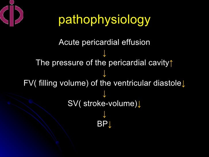
Pericardial diseases can present clinically as acute pericarditis, pericardial effusion, cardiac tamponade, and constrictive pericarditis. Patients can subsequently develop chronic or recurrent pericarditis. Structural abnormalities including congenitally absent pericardium and pericardial cysts are usually asymptomatic and are uncommon. Clinicians are often faced with several …
Pleural effusion; Diagram of fluid buildup in the pleura: Specialty: Pulmonology: A pleural effusion is accumulation of excessive fluid in the pleural space, the potential space that surrounds each lung.Under normal conditions, pleural fluid is secreted by the parietal pleural capillaries at a rate of 0.01 millilitre per kilogram weight per hour, and is cleared by lymphatic …
Extensive reviews on the pathophysiology of pleural effusions are available 100-104. The causes of pleural effusion may be subdivided into three main categories: 1) those changing transpleural pressure balance, 2) those impairing lymphatic drainage, and 3) those producing increases in mesothelial and capillary endothelial permeability.
Pleural Effusion By Richard W. Syndrome occurring as a effusion pathophysiology diagram of ovulation induction with human chorionic gonadotropin hCG and occasionally hypothyroidsim. Chylous effusion chylothorax is a milky white effusion high in triglycerides caused by traumatic or neoplastic most often lymphomatous damage to the thoracic duct.
A pleural effusion is collection of fluid abnormally present in the pleural space, usually resulting from excess fluid production and/or decreased lymphatic absorption. [] It is the most common manifestation of pleural disease, and its etiologies range in spectrum from cardiopulmonary disorders and/or systemic inflammatory conditions to malignancy.
pathophysiology pleural effusion diagram. buy aspira pleural drainage. biapical nodular pleural scarring. mild basilar pleural thickening trauma. left apical pleural thickening. pictures of children postures. mild apical pleural. right pleural friction rub. mild bilateral apical pleural parenchymal scarring. edema in pleural cavity nodules
A malignant pleural effusion (MPE) is the build up of fluid and cancer cells that collects between the chest wall and the lung. This can cause you to feel short of breath and/or have chest discomfort. It is a fairly common complication in a number of different cancers.








_CRUK_054.svg/1200px-Diagram_showing_a_build_up_of_fluid_in_the_lining_of_the_lungs_(pleural_effusion)_CRUK_054.svg.png)


0 Response to "35 pleural effusion pathophysiology diagram"
Post a Comment