40 diagram of lower back muscles
We are pleased to provide you with the picture named Anatomy Of Back Muscles Diagram.We hope this picture Anatomy Of Back Muscles Diagram can help you study and research. for more anatomy content please follow us and visit our website: www.anatomynote.com. Anatomynote.com found Anatomy Of Back Muscles Diagram from plenty of anatomical pictures on the internet. Muscles Of The Lower Back And Buttocks Diagram. Hold for 2 to 3 seconds, inhale, and slowly lower yourself back to your starting position. The gluteal muscle group is comprised of three muscles, gluteus maximus, gluteus minimus, and gluteus medius. Nerves that run through and . The sciatic nerve is located inferiorly to this muscle, so if the .
The muscles of the lower back help stabilize, rotate, flex, and extend the spinal column, which is a bony tower of 24 vertebrae that gives the body structure and houses the spinal cord.The spinal ...

Diagram of lower back muscles
The muscles of the back can be arranged into 3 categories based on their location: superficial back muscles, intermediate back muscles and intrinsic back muscles.The intrinsic muscles are named as such because their embryological development begins in the back, oppose to the superficial and intermediate back muscles which develop elsewhere and are therefore classed as extrinsic muscles. Pelvic floor muscles and ligaments attach to the coccyx. What conditions and disorders affect the spine? Up to 80% of Americans experience back ... The abdominal and lower back muscles work together to form a supportive "girdle" around your waist and lower back. Stronger muscles can help stabilize the lower back and can help reduce injury risk. 3. Stop smoking. Nicotine reduces blood flow to the spinal structures, including the lumbar discs, and can accelerate age-related degenerative ...
Diagram of lower back muscles. For more complete coverage of the structure and function of the low back and pelvis, The Muscular System Manual – The Skeletal Muscles of the ... See Back Muscles and Low Back Pain. Nerves in your lower back. Five pairs of lumbar spinal nerves labeled L1 to L5 branch off your spinal cord and exit through small holes between the vertebrae. The part of the nerve that emerges out of the spine is called the nerve root. Lower Back Muscles Diagram Diagram Of Lower Back Muscles Human Anatomy Diagram, Picture of Lower Back Muscles Diagram Diagram Of Lower Back Muscles Human Anatomy Diagram Muscle Problems and Low Back Pain. Most low back pain is caused by muscle strain or a sprain. A strain is when your muscle fibers are stretched. A sprain is when your ligaments (bands of tissue ...
Keep your back muscles in good shape to prevent back pain. Exercises that strengthen the core (abdominals and lower back) can help to protect the spine from damage. When sitting at a desk, watch your posture and get up to stretch your legs every 20 minutes to an hour. Use proper form when lifting heavy objects—lift from your legs, not your back. The muscles of the lower back help stabilize, rotate, flex, and extend the spinal column, which is a bony tower of 24 vertebrae that gives the body structure related searches for muscle diagram labeled back diagram of back muscles painanatomy of back muscles imageback muscles. The back consists of the spine, spinal cord, muscles, ligaments, and nerves. These structures work together to support the body, enable a range of movements, and send messages from the brain to ... Bones of the Pelvis and Lower Back. The bones of the pelvis and lower back work together to support the body's weight, anchor the abdominal and hip muscles, and protect the delicate vital organs of the vertebral and abdominopelvic cavities. The vertebral column of the lower back includes the five lumbar vertebrae, the sacrum, and the coccyx.
25,583 back muscle anatomy stock photos, vectors, and illustrations are available royalty-free. See back muscle anatomy stock video clips. of 256. human musculature bodybuilding infographic muscular system vector human anatomy back muscle anatomy bicep male muscular anatomy human body anatomy female female anatomy muscle hamstrings muscle ... Anterior and posterior muscles of the lower arm ... Client Back Care Guide. FREE Download. Pain-free clients are happy clients. Claim your free copy of the client back care guide today. Your clients will thank you for it! Link to Client Back Care Guide. Links to FMA Sales Page. Lower Back Pain Exercises. Back muscles, like any other muscle in the body, require adequate exercise to maintain strength and tone. See How Exercise Helps the Back. While muscles like the gluteals (in the thighs) are used any time we walk or climb a step, deep back muscles and abdominal muscles are usually not actively engaged during everyday activity. The fascia covers and supports the muscles. Back Muscles. Your back muscles extend from the bones of your neck (cervical vertebrae) to your lower back (lumbar spine) and then to the base of your lumbar spine (sacrum) and tailbone (coccyx). Some of these muscles are quite large and cover broad areas, e.g. large areas of the trunk.

Gene and Nancy Bavinger House: Plan of Lower Level Showing Water Garden (1950/51) // Bruce Alonzo Goff American, 1904–1982
Most superficial is the strong Erector Spinae or sacrospinalis muscle. · Middle layer is the multifidus. · Third layer is made up of small muscles arranged from ...
Deep back muscles. The deep back muscles, also called intrinsic or true back muscles, consist of four layers of muscles: superficial, intermediate, deep and deepest layers.These muscles lie on each side of the vertebral column, deep to the thoracolumbar fascia They span the entire length of the vertebral column, extending from the cranium to the pelvis
Muscles of the Low Back and Pelvis - posterior superficial view Muscles of the Low Back and Pelvis - right lateral deep view Muscles of the Low Back and Pelvis - right lateral superficial view
Jan 6, 2020 - Explore Abdullah Shekh's board "Lower back muscles anatomy" on Pinterest. See more ideas about muscle anatomy, anatomy, human anatomy and physiology.
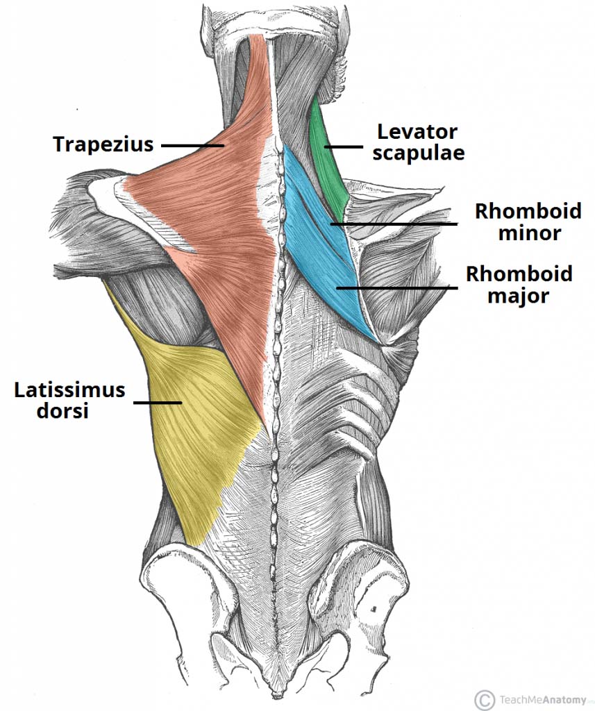
Back anatomy. The back is the body region between the neck and the gluteal regions. It comprises the vertebral column (spine) and two compartments of back muscles; extrinsic and intrinsic. The back functions are many, such as to house and protect the spinal cord, hold the body and head upright, and adjust the movements of the upper and lower limbs.
The back has three natural curves which form an S-shape when viewed from the side. This helps to balance and distribute the weight of the body on the legs. Cervical Spine Diagram Stress in the spine is greatest in the cervical (neck) and lumbar (lower back) areas. These two regions are responsible for most of the movement in the back, allowing ...
Back Muscle Diagram With Lower Back Anatomy. This is a diagram of the larger and more surface muscles of the low back. The anatomy of the spine is complicated. To learn more, watch this VIDEO. Lower Back Muscle Diagram Anatomy Does Degenerative Disc Disease affect the Lower Back Muscles? Another common cause of lower back and hip pain is disc.
The four muscle groups that together make up the deep muscle group are the segmental muscles, the transversospinales, the erector spinae, and the spinotransversales. "The only way to isolate specific back muscles - whether it is upper or lower back - or make any progress is through the power of the mind-muscle connection."
The Quadratus Lumborum (QL) muscle, as seen in the diagram above, has a vital role in side bending your spine. The QL is a deep muscle that runs on both sides of the lower back. The muscle begins on the lowest rib (rib 12) and the nearby vertebra and connects to the pelvic crests.

Image from page 144 of "How to keep well : a text-book of health for use in the lower grades of schools with special reference to the effects of alcholic drinks, tobacco and other narcotics on the bodily life" (1901)
Muscles Of Lower Back Diagram. In this image, you will find an occipital bone, sternocleidomastoid, trapezius, deltoid in Muscles of the lower back diagram. As you can see, there are also have a spine of scapula deltoid, triceps brachii, latissimus dorsi. This picture also contains humerus, olecranon process of ulna, deep to tendon and so on.
Deep muscles of the lower back include: The multifidus, a long muscle that travels nearly the entire length of the back.It helps to stabilize and rotate the lower back, and additionally takes some ...
Lumbar (lower back) muscle strains and sprains are the most common causes of low back pain. Muscle strains and sprains are common in the lower back, because it supports the weight of the upper body and is involved in moving, twisting and bending. Lumbar muscle strain is caused when muscle fibers are abnormally stretched or torn.
Symptoms of Low Back Pain. 2 / 25. These might range from a dull ache to a stabbing or shooting sensation. The pain may make it hard to move or stand up straight. Pain that comes on suddenly is ...
The muscles of the abdomen, lower back, and pelvis are separated from those of the chest by the muscular wall of the diaphragm, the critical breathing muscle. Lying exposed between the protective bones of the superiorly located ribs and the inferiorly located pelvic girdle, the muscles of this region play a critical role in protecting the ...
The abdominal and lower back muscles work together to form a supportive "girdle" around your waist and lower back. Stronger muscles can help stabilize the lower back and can help reduce injury risk. 3. Stop smoking. Nicotine reduces blood flow to the spinal structures, including the lumbar discs, and can accelerate age-related degenerative ...
Pelvic floor muscles and ligaments attach to the coccyx. What conditions and disorders affect the spine? Up to 80% of Americans experience back ...
The muscles of the back can be arranged into 3 categories based on their location: superficial back muscles, intermediate back muscles and intrinsic back muscles.The intrinsic muscles are named as such because their embryological development begins in the back, oppose to the superficial and intermediate back muscles which develop elsewhere and are therefore classed as extrinsic muscles.

Amulet of the Crown of Lower Egypt (Third Intermediate Period, Dynasty 21–25 (1070–656 BCE)) // Egyptian


0 Response to "40 diagram of lower back muscles"
Post a Comment