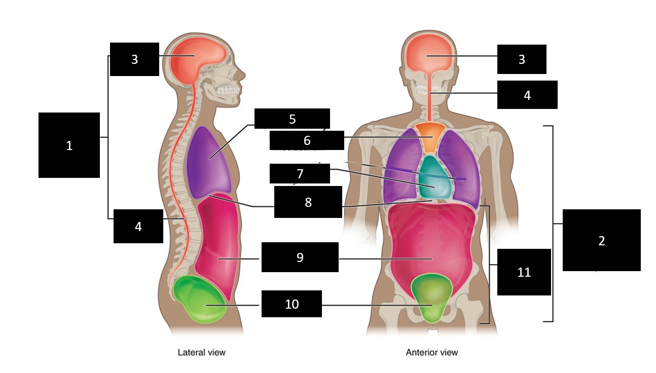
37 ventral body cavity diagram
External View, Ventral Nerve Cord and Ventral Blood Vessel Labeled Nerve Cord & Blood Vessel; Note the ventral nerve cord with segmental ganglia (ganglion = a knot on a string). If you lift a section of the digestive tract/blood vessels, the nerve cord should be seen lying on the ventral surface of the body cavity. Greater omentum (ventral view) On the other hand, the ventral mesentery suspends only the lower esophagus, the stomach, and the upper duodenum to the ventral body wall.The ventral mesentery is derived from the septum transversum (a mass of splanchnic mesoderm separating the pericardial and peritoneal cavities) and eventually becomes divided by the growing liver into the lesser omentum and the ...
The circulatory system is an open type in Hemichordata, circulatory fluid is colorless, respiratory pigment is absent. A dorsal heart pumps blood throughout the body and there are two main blood vessels, one dorsal vessel on the dorsal side and another one is the ventral vessel on the ventral side. Excretion in Hemichordata:

Ventral body cavity diagram
The pelvic cavity is a bowl-like structure that sits below the abdominal cavity. The true pelvis, or lesser pelvis, lies below the pelvic brim (Figure 1). This landmark begins at the level of the sacral promontory posteriorly and the pubic symphysis anteriorly. The space below contains the bladder, rectum, and part of the descending colon. In females, the pelvis also houses the uterus ... IMAIOS and selected third parties, use cookies or similar technologies, in particular for audience measurement. Cookies allow us to analyze and store information such as the characteristics of your device as well as certain personal data (e.g., IP addresses, navigation, usage or geolocation data, unique identifiers). The ventral body is modified into a muscular plough-like surface, the foot which is variously modified for creeping, burrowing and swimming. Mantle or pallium is a fold of a body wall that leaves between itself the main body, mantle cavity.
Ventral body cavity diagram. Dogfish shark dissection diagram. List of the 5 fins of the dogfish shark 2 dorsal pectoral pelvic caudal the depressor of the pectoral fin allows the pectoral fins to lower. Fix the specimen on the tray keeping the ventral surface up by pushing pins through the fins and if necessary also through the muscles of the lateral body wall. Visceral fat is a type of body fat that s stored within the abdominal cavity. Any empty or hollow space within the body a hole or hollow space in an organ tissue or other body part. Anat the ventral cavity of a vertebrate which contains the alimentary canal as distinguished from the dorsal or cerebro spinal canal. Chapter 1: Atomic position. The Orbital Cavities House The Right Hypochondriac Region Major Body Cavities Human Body Temperature Right Lumbar Region. TERMS IN THIS SET (39) Identify the cavity that houses the spinal cord. For this diagram it's 3. Identify the cavity that houses the heart and lungs ( and other organs) j. Dorsal body cavity–the cranial cavity and the spinal cavity in combination. · Cranial cavity–the space occupied by the brain, enclosed by the skull bones.
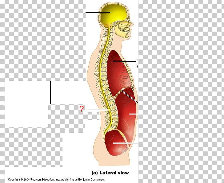
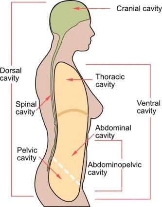
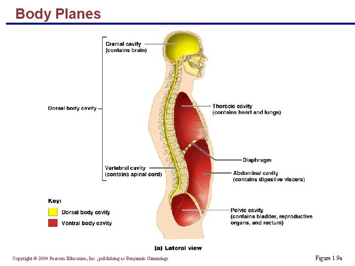
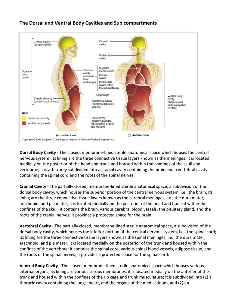
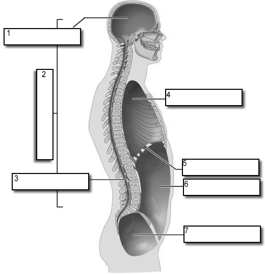
The coronal plane or frontal plane divides the body into dorsal and ventral portions whereas the transverse plane or axial plane divides the body into cranial and caudal portions. Diagram of the Human Body. For each organ system identify some of its organs and functions. Enzymes help speed up chemical reactions in the body. Another name for anterior is ventral, and another name for posterior is dorsal. For ventral, you can think of a ventriloquist which literally translates to "stomach talker". We learned in our medical prefix lecture that the prefix "ventri-" refers to the stomach, abdomen, or front of the body. This can help you remember ventral means ... Cranial cavity-the space occupied by the brain, enclosed by the skull bones. Spinal cavity-the space occupied by the spinal cord enclosed by the vertebrae column making up the backbone. The spinal cavity is continuous with the cranial cavity. Ventral body cavity-the thoracic cavity, the abdominal cavity, and the pelvic cavity in combination. Respiratory System Unlabeled Human Anatomy Diagram Coloring Home. 2 Representative diagram of the relationship between viscera coelomic cavity and the body wall. The ventral body chamber that contains the. In mammals the diaphragm separates the thoracic cavity from the abdominal cavity. The spinal cavity is continuous with the cranial cavity.
Start studying Body Cavities anterior View labeling. Learn vocabulary, terms, and more with flashcards, games, and other study tools. Scheduled maintenance: Saturday, December 12 from 3-4 PM PST Body cavities in the human body.Human body organ diagram front view. The dorsal body cavity protects organs of the nervous system and has two subdivisions. The cranial cavity is the area within the skull and encloses the brain. The medullary cavity contains red bone marrow during childhood eventually turning into yellow bone marrow after puberty. Ventral body cavity — The dorsal body cavity includes the cranial cavity, enclosed by the skull and contains the brain, and the spinal cavity, enclosed by ...Human body cavities · Development · Function · Other animals The abdominopelvic cavity is the area that houses the organs beneath the lungs and heart. Learn the nine regions of this area, and the features of bone and organs as well.
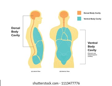
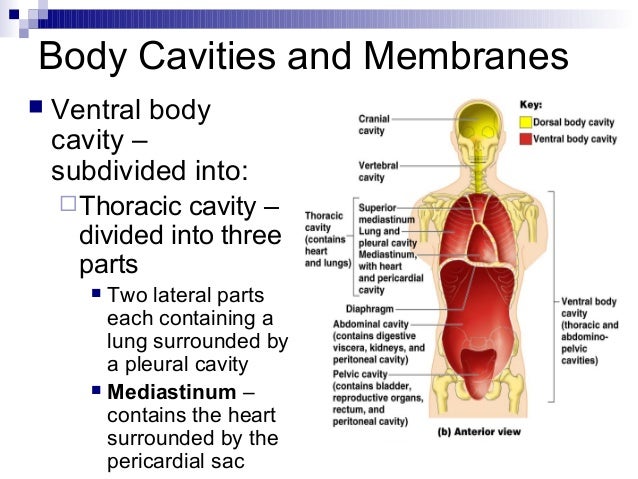
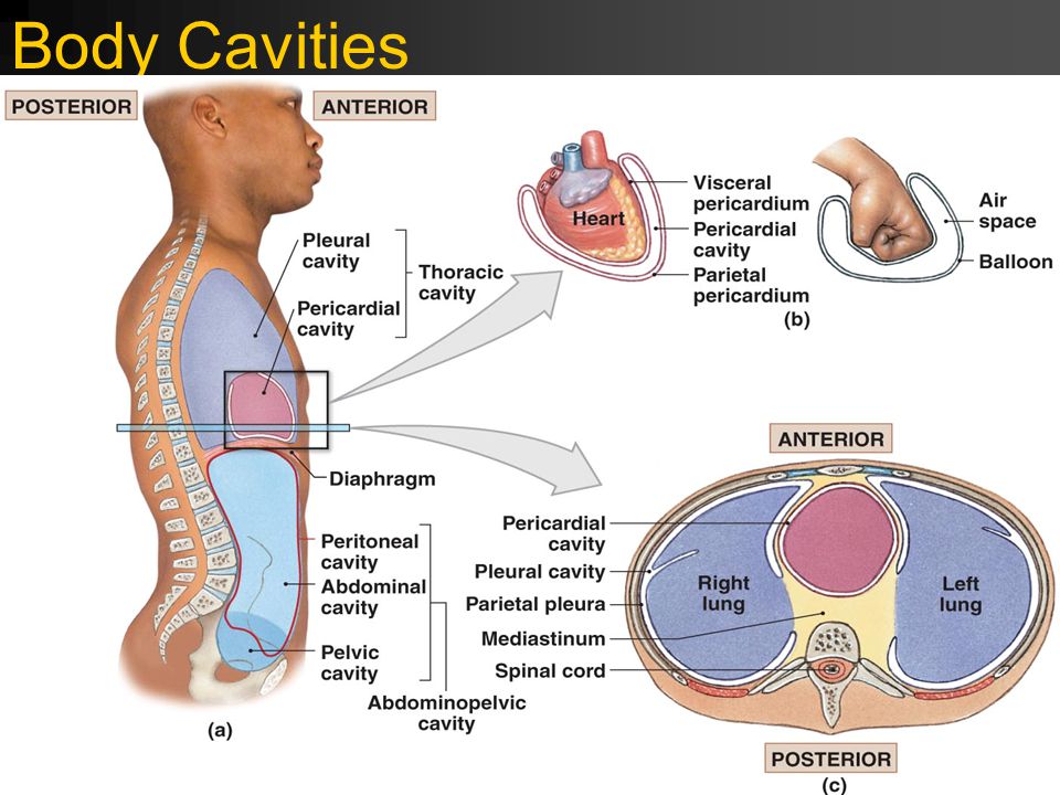
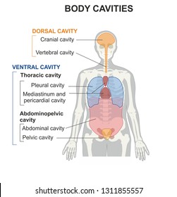
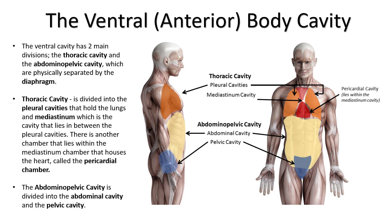
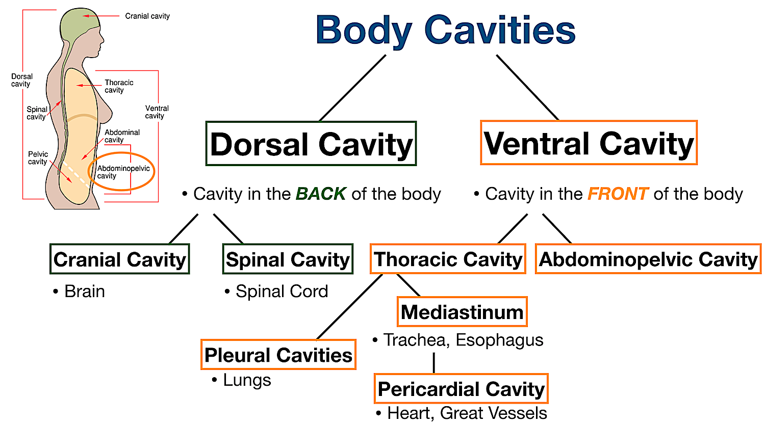
The ventral body cavity is a human body cavity that is in the anterior aspect of the human body. It is made up of the thoracic cavity , and the abdominopelvic cavity . The abdominopelvic cavity is further divided into the abdominal cavity and pelvic cavity, but there is no physical barrier between the two.
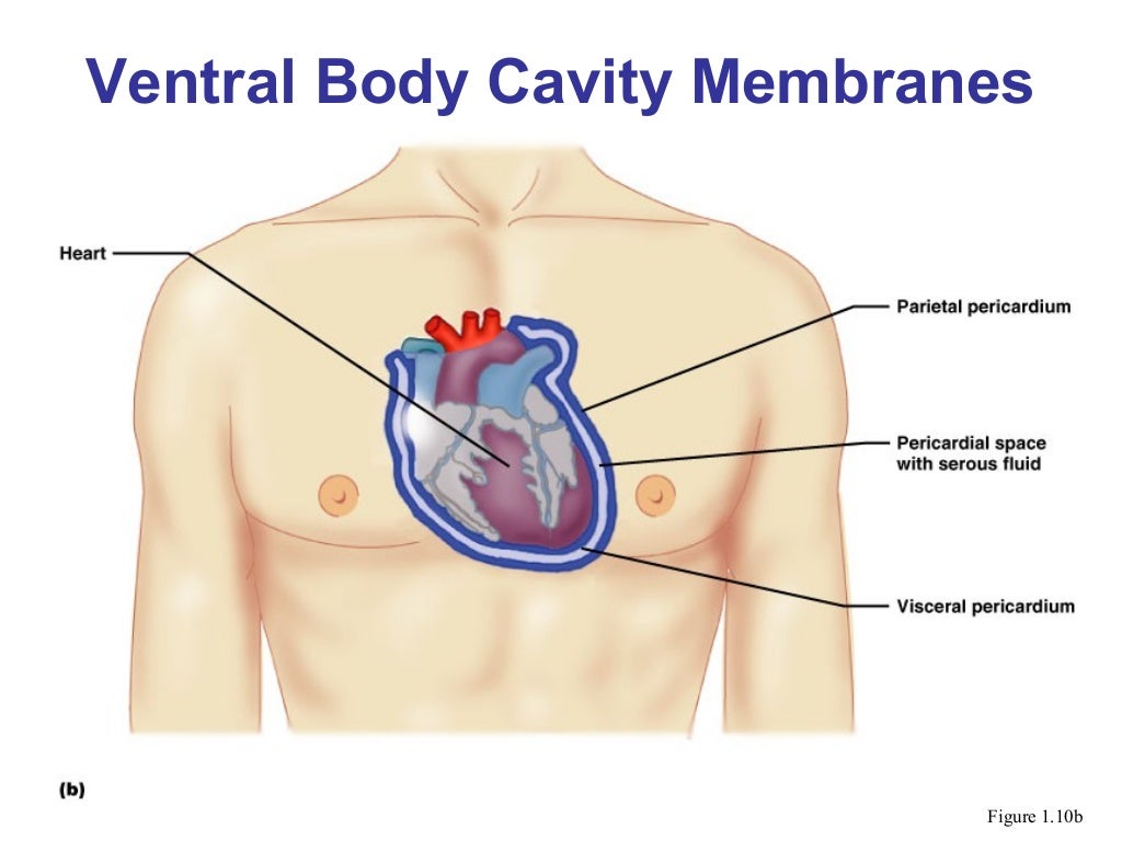
Ventral — The dorsal cavity contains the primary organs of the nervous system, including the brain and spinal cord. The diaphragm is a sheet of muscle that ...
The body of the bird ischium bone contributes to forming the acetabulum cavity caudally. It also contributes to forming the part of the ventral antitrochanter. The wing of the bird ischium bone locates at the lateral position. It forms the ventral continuation of the lateral surface of the ilium bone.
Buccal Cavity: The mouth opens into a cavity, buccal cavity and this portion ends at the collar-trunk septum. ... The sacculation is conspicuous from outside of the body wall in the hepatic region which is known as hepatic caeca. The posthepatic region of the intestine is remain touched with the ventral body wall and the Pygochord is present ...

Endocrine system diagram worksheet. Worksheet 2.6 kinematic equations answer key Body Cavities. The cavities, or spaces, of the body contain the internal organs, or viscera.The two main cavities are called the ventral and dorsal cavities. The ventral is the larger cavity and is subdivided into two parts (thoracic and abdominopelvic cavities) by ...

Human internal organs dummy training dummy detail of the uscular system. A The dorsal cavity. Gallbladder disease is the name used to several conditions that can impact your gallbladder. Diagram of the Digestive System And an Explanation of its Working. Body cavities hollow spaces within the human body that contain internal organs.
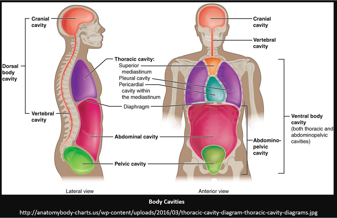
Body Cavities Labeled Diagram: The ventral cavity is located in the front of the body (red/star) and houses the organs/structures of the chest, abdomen, and pelvis. Cranial Cavity Now that we have a good understanding of the dorsal and ventral cavities, each cavity can be broken down even more.
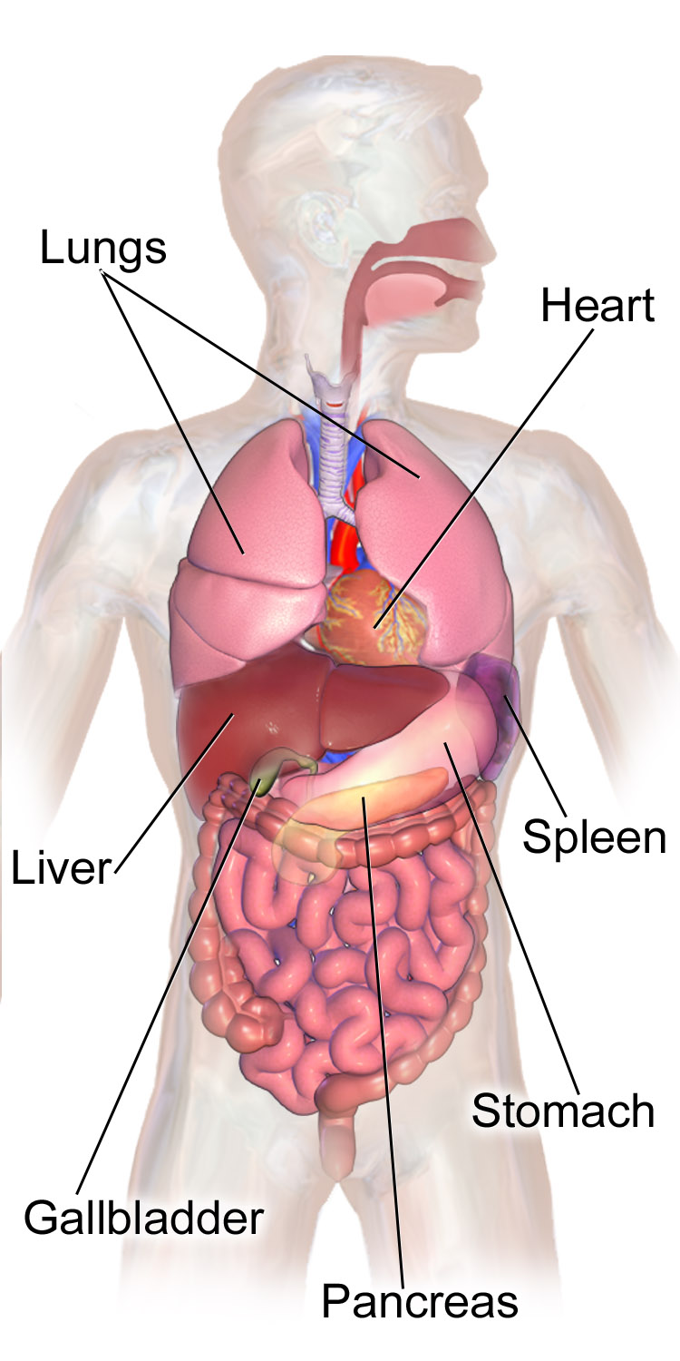
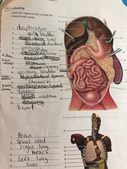
The ventral cavity contains the heart, lungs, trachea, esophagus, stomach, liver, pancreas, gall bladder, spleen, kidneys, small and large ...5 Sept 2021What organs are in the ventral cavity?Is the diaphragm in the ventral cavity?
A body cavity is a fluid-filled space inside the body that holds and protects internal organs. Human body cavities are separated by membranes and other structures. The two largest human body cavities are the ventral cavity and the dorsal cavity. These two body cavities are subdivided into smaller body cavities.

- Gut tube & body cavity -tube on tube and formation of body cavity to be read and concept grip - Ventral body wall defects* do all - Diaphragm formation v.imp+diagrams and diaphragmatic hernia -Axial skeleton

Body cavities diagram from an animal. You already got a different diagram of the body cavity from an animal. Here, I will show you again some of the cavities in one diagram. But, if you need more updated diagrams on the body cavity, you may join anatomy learner on social media.

Q.1. Which is the longest bone of our body? Ans: Femur or thigh bone is the longest bone of our body. Q.2. What are the functions of the axial skeleton? Ans: The axial skeleton provides support and protection for the brain, spinal cord, and the organs in the ventral body cavity. Q.3. What is the role of the appendicular skeleton?

Thoracic wall The first step in understanding thorax anatomy is to find out its boundaries. The thoracic, or chest wall, consists of a skeletal framework, fascia, muscles, and neurovasculature - all connected together to form a strong and protective yet flexible cage.. The thorax has two major openings: the superior thoracic aperture found superiorly and the inferior thoracic aperture ...
Anatomical directional terminology concerns the location of appendages on the human body. Learn about anatomical directional terminology used for the limbs, hands, and feet, and explore the ...
Acquire the dogfish shark dissection diagram study guide connect that we give here and check out the link. The olfactory bulbs are. Crab ...

The ventral body is modified into a muscular plough-like surface, the foot which is variously modified for creeping, burrowing and swimming. Mantle or pallium is a fold of a body wall that leaves between itself the main body, mantle cavity.

IMAIOS and selected third parties, use cookies or similar technologies, in particular for audience measurement. Cookies allow us to analyze and store information such as the characteristics of your device as well as certain personal data (e.g., IP addresses, navigation, usage or geolocation data, unique identifiers).
The pelvic cavity is a bowl-like structure that sits below the abdominal cavity. The true pelvis, or lesser pelvis, lies below the pelvic brim (Figure 1). This landmark begins at the level of the sacral promontory posteriorly and the pubic symphysis anteriorly. The space below contains the bladder, rectum, and part of the descending colon. In females, the pelvis also houses the uterus ...


0 Response to "37 ventral body cavity diagram"
Post a Comment