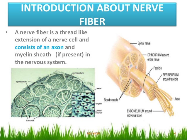
36 which part of the diagram is considered nerve fiber
Tear production is influenced by parasympathetic fibers in the facial nerve, which activate a ganglion, and ultimately the lacrimal (tear) gland. Neurons in the dorsal nucleus of the vagus nerve and the nucleus ambiguus project through the vagus nerve (cranial nerve X) to the terminal ganglia of the thoracic and abdominal cavities ... Which part of the diagram is considered nerve fiber? A D I Both A and D All of these choices make up the nerve fiber. Both A and D. Na+/K+-ATPase is considered to be an electrogenic pump because A. it contributes to the negativity of the resting membrane potential.
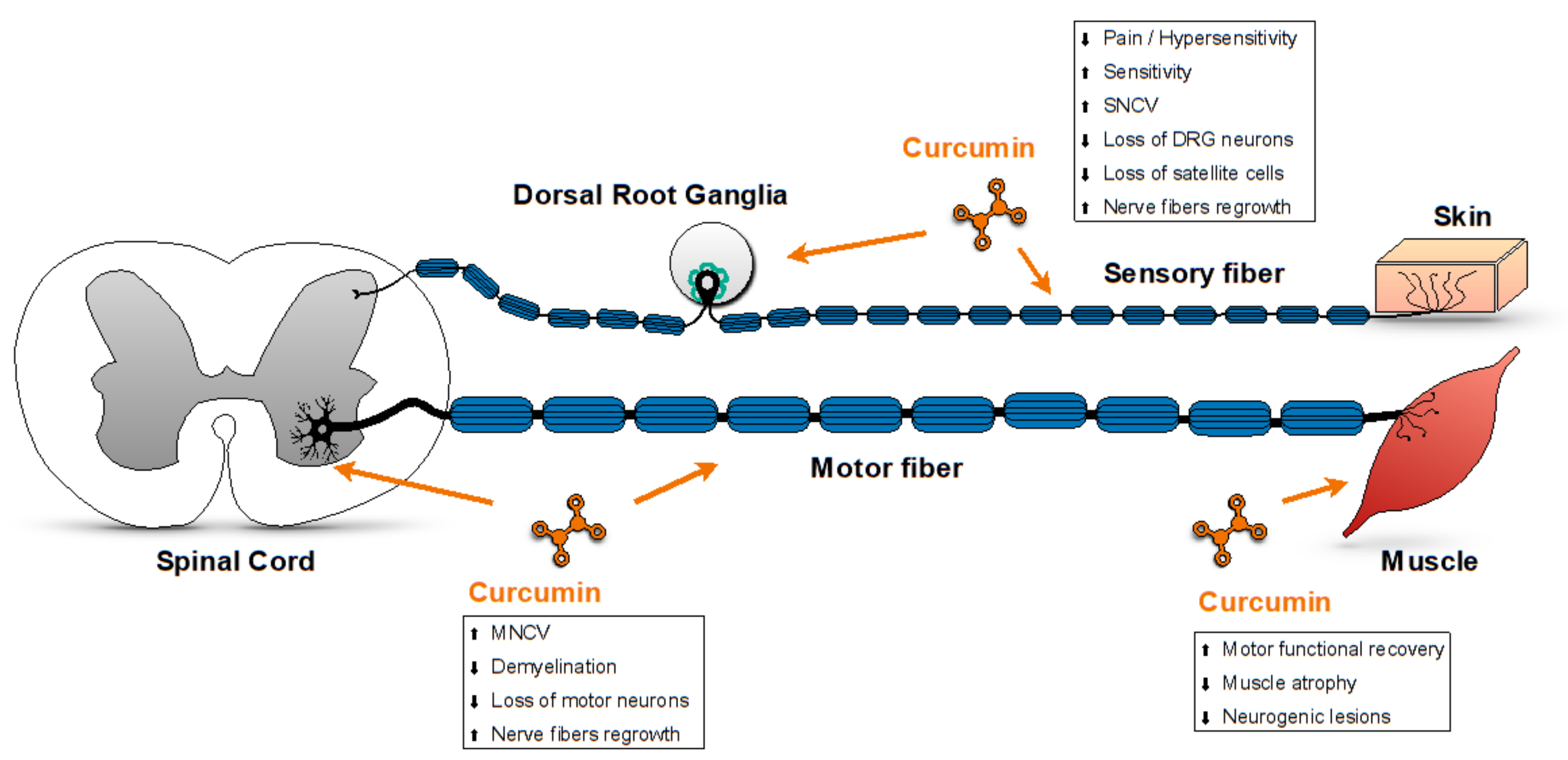
Nov 08, 2021 · Sympathetic nerve fibers sprout into DRGs after peripheral nerve injury (A) Diagram showing the mating strategy. GFP is expressed in DRG neurons, tdT is expressed in the sympathetic axons. (B–D) Whole-mount cleared L4 DRGs of mice with and without SNI (day 21). (B0–B2) Naive mouse. (B3–B5) SNI mouse.
Which part of the diagram is considered nerve fiber
R.A. Armstrong, in The Neuroscience of Cocaine, 2017 1.5.4 Visual System. The nerve fibers of the retina, which represent the axons of the ganglion cells, collect together at the optic disk and leave the eye as the optic nerve, subsequently passing into the brain through the orbital bones. The optic nerves from each eye meet at the optic chiasm, a structure at the base of the brain where nerve ... In order to contract, skeletal muscle fibers must be voluntarily stimulated by the nervous system. The site of muscle stimulation, where the nerve fiber communicates with the muscle fiber, is called the neuromuscular junction. Aug 16, 2012 · The syndrome is placed in this portion of the diagram, not because it is due to lesions located in this area, but rather because lesions located elsewhere intracranially can secondarily affect the sixth nerve; this is an example of the poor localizing value of a sixth nerve palsy.
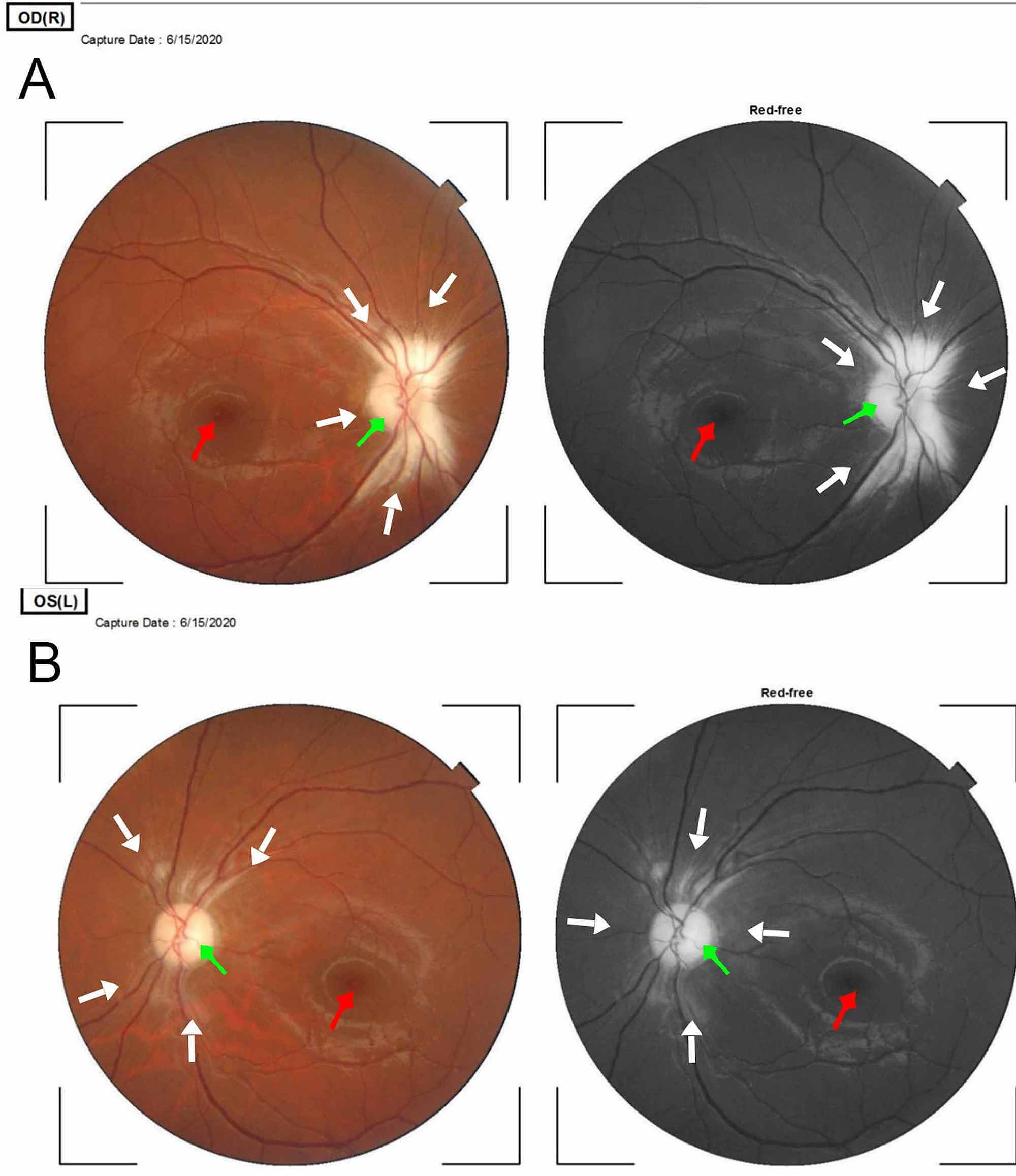
Which part of the diagram is considered nerve fiber. Nerve fiber refers to: a. Axon. b. Dendrites. c. Nissl body. d. Both a and b. e. All of the above. D. 7 ... Sodium pumps are considered electrogenic because. a. They contribute to the negativity of the resting membrane potential ... Which part of the diagram is considered nerve fiber? a. A b. D c. H d. Bothaandb e. All of the above. E. Decks in ... Structure. Each gap junction (aka nexus junction) contains numerous gap junction channels that cross the plasma membranes of both cells. With a lumen diameter of about 1.2 to 2.0 nm, the pore of a gap junction channel is wide enough to allow ions and even medium-size molecules like signaling molecules to flow from one cell to the next, thereby connecting the two cells' cytoplasm. Which part of the diagram is considered nerve fiber? A, B. This part of the diagram contains organelles and Nissl bodies. E. This portion of the diagram contains cytoplasm and a myelin sheath wrapped around neurolemma. C. In the diagram, where is the axon collateral? H. Temporal to the optic nerve head at a distance approximately 2.5 optic nerve (disk) diameters at roughly 3.4 mm distance lies a dark brown-yellowish area (Figure 1), in the center of which is the tiny circular fovea. The position of the fovea can be seen clearly in the retina illustrated in Figure 2A.
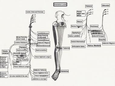
Section Reference 1: Sec 12.2 Histology of Nervous Tissue. 36) This structure electrically insulates the axon of a neuron to increase the speed of nerve impulse conduction. a) A b) Bc) C d) D e) E Answer: e. Difficulty: EasyStudy Objective 1: SO 12.2 Compare the structures and functions of neurons and neuroglia and white matter and gray matter. Saphenous Nerve It is the largest cutaneous branch of the femoral nerve. It supplies cutaneous branches to the skin of the leg and foot in the region between the knee and the ankle. Sciatic Nerve Also known as the ischiatic nerve, the sciatic nerve is a nerve fiber that begins in the lower back and ends in the lower limb. View ch12.docx from BIOLOGY 235 at Athabasca University, Athabasca. Package Title: Testbank Course Title: pap14 Chapter Number: 12 1) Which of the following is NOT a function of the nervous Which part of the diagram is considered nerve fiber? a) A b) D c) I d) Both A and D e) All of these choices make up the nerve fiber. D. This part of the neuron contains the nucleus and Nissl bodies. a) A b) B c) C d) E e) Both A and B. B.
These nerve fibers are considered spinal nerves, which carry and transmit information between the spinal cord and parts of the body. The nerves stem from portions of the vertebrae. May 15, 2018 · Root. The root is the part of the tooth that extends into the bone and holds the tooth in place. It makes up approximately two-thirds of the tooth. Which part of the diagram is considered nerve fiber? a. A b. D c. H d. Both a and b e. All of the above. e. All of the above. Refer to image 1 37. This part of the neuron contains the nucleu and Nissl bodies. a. A b. B c. C d. E e. Both a and b. b. B. Refer to image 1 38. This part of a neuron contains cytoplasm and a myelin sheath wrapped ... Which part of the diagram is considered nerve fiber. Related diagrams and images facial nerve. Refer to image 1 37. 69 the function of this pathway is to convey nerve impulses from the brainstem to cause automatic movements that regulate muscle tone posture and balance and orientation of the head and body.
Which part of the diagram is considered nerve fiber? a. A b. D c. I d. Both A and D e. All of these choices make up the nerve fiber. 46. In the diagram, where is a node of Ranvier? a. A b. B c. C d. D e. E. 47. Which of the channels shown in the figure opens and closes randomly? a. A b. B c. C d. D e.

Oct 10, 2021 · A single nerve impulse of a motor neuron will cause a motor unit to contract briefly before relaxing. This small contraction is known as a twitch contraction. If the motor neuron provides several signals within a short period of time, the strength and duration of the muscle contraction increases.

1) Which part of the diagram is considered nerve fiber? a) A b) D c) I d) Both A and D e) All of these choices make up the nerve fiber

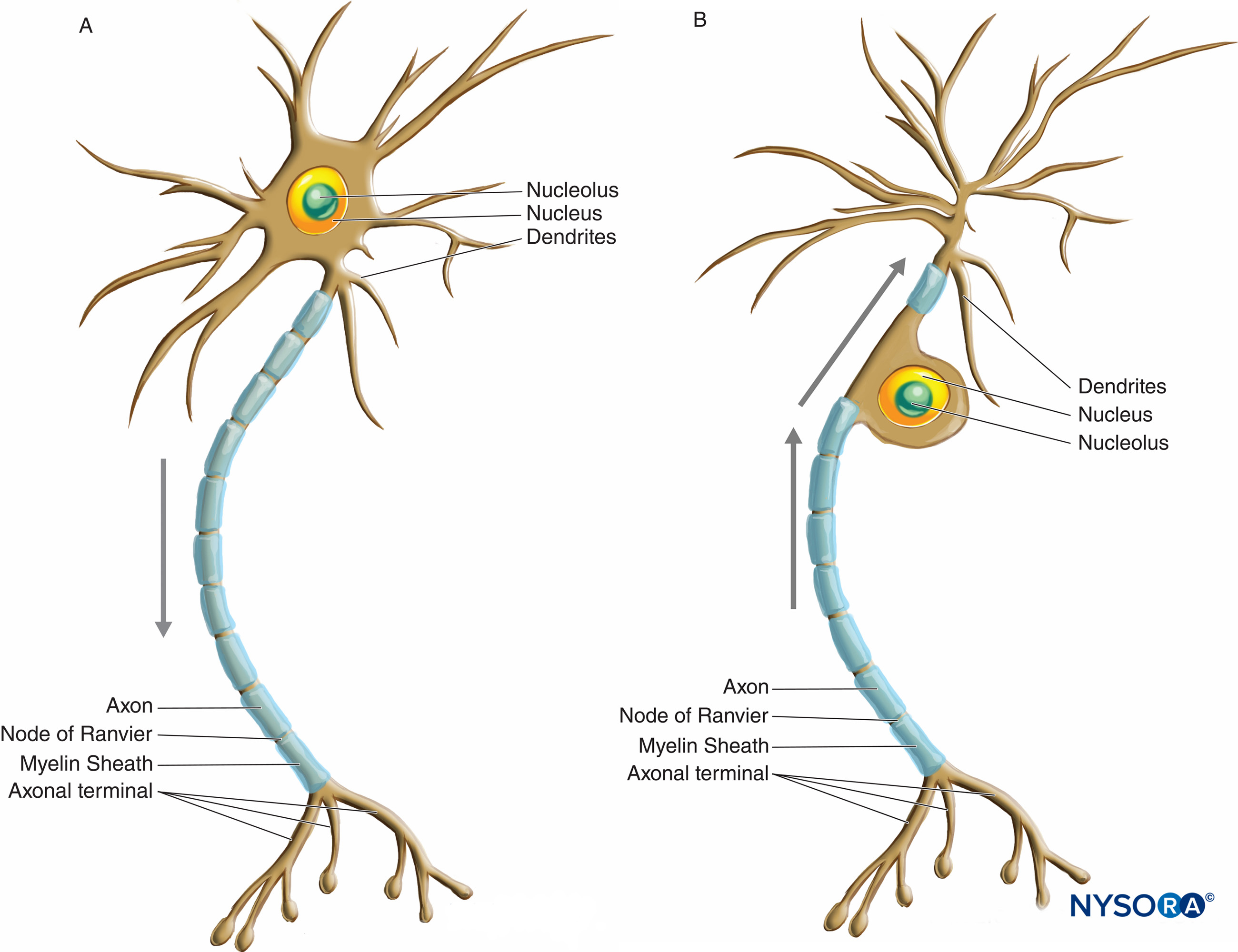
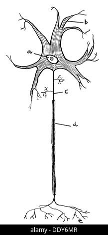
Parts of the Nerve Cell and Their Functions Silvia Helena Cardoso, PhD [1. Cell body] [2.Neuronal membrane][3.Dendrites] [4. Axon][5. Nerve ending] 1. Cell body The (soma) is the factory of the neuron. It produces all the proteins for the dendrites,axons and synaptic terminals and contains specialized organelles such asthe mitochondria, Golgi ...

Which part of the diagram is considered nerve fiber? A)A B)D C)I D)Both A and D E)All of these choices make up the nerve fiber. Free. Multiple Choice . Unlock to view answer. Q 37 Q 37. This part of the neuron contains the nucleus and Nissl bodies. A)A B)B C)C D)E E)Both A and B. Free.
Which part of the diagram is considered nerve fiber? a) Dendrites b) Cell body c) Axon collateral d) Axon e) Neurolemma f) Axoplasm g) Axolemma h) Axon terminal i) Synaptic end bulb Both A and D. This part of the neuron contains the nucleus and Nissl bodies. a) Dendrites b) Cell body c) Axon collateral d) Axon
69) The function of this pathway is to convey nerve impulses from the brainstem to cause automatic movements that regulate muscle tone, posture, and balance and orientation of the head and body. a) Indirect pathway

Which part of the neuron in this diagram produces proteins that can regenerate damaged axons in the PNS? Which part of the neuron in this diagram will contain voltage-gated ion channels? In this diagram, which structure electrically insulates the axon of a neuron to increase the speed of nerve impulse conduction?
Which part of the diagram is considered nerve fiber? a) A b) D c) I d) Both A and D e) All of these choices make up the nerve fiber. d. In the diagram, where are axon terminals? a) F b) G c) H d) I e) None of these choices. c) H. This part of the neurons contains the nissl bodies. a) A b) B c) C d) D e) both A and B. B.
Aug 16, 2012 · The syndrome is placed in this portion of the diagram, not because it is due to lesions located in this area, but rather because lesions located elsewhere intracranially can secondarily affect the sixth nerve; this is an example of the poor localizing value of a sixth nerve palsy.
In order to contract, skeletal muscle fibers must be voluntarily stimulated by the nervous system. The site of muscle stimulation, where the nerve fiber communicates with the muscle fiber, is called the neuromuscular junction.
R.A. Armstrong, in The Neuroscience of Cocaine, 2017 1.5.4 Visual System. The nerve fibers of the retina, which represent the axons of the ganglion cells, collect together at the optic disk and leave the eye as the optic nerve, subsequently passing into the brain through the orbital bones. The optic nerves from each eye meet at the optic chiasm, a structure at the base of the brain where nerve ...


0 Response to "36 which part of the diagram is considered nerve fiber"
Post a Comment