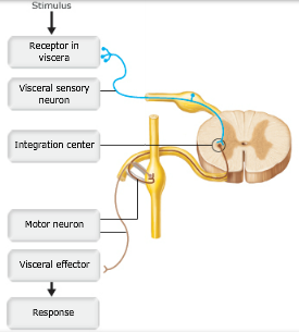
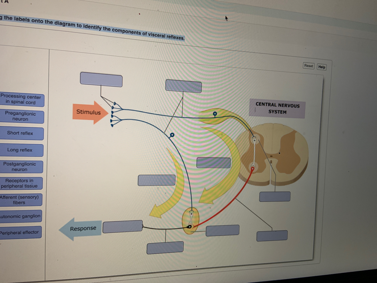
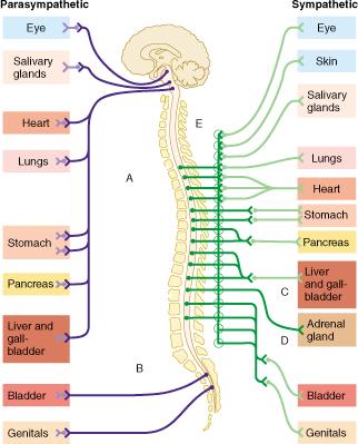
38 drag the labels onto the diagram to identify the components of the autonomic nervous system.
The peripheral nervous system is made up of thick bundles of axons, called nerves, carrying messages back and forth between the CNS and the muscles, organs, and senses in the periphery of the body (i.e., everything outside the CNS). The PNS has two major subdivisions: the somatic nervous system and the autonomic nervous system. Drag the labels onto the diagram of neurochemical communication at an autonomic synapse. ANSWER: Correct. Art-labeling Activity Figure 11. Label the parts of the neuromuscular junction. Part A. Drag the labels onto the diagram to identify parts of the neuromuscular junction. ANSWER: Reset Help. Action potential arrives at varicosity.
autonomic nervous system. Prag the labels onto the diagram to identify the components of the autonomic nervous system! Reset Help Cardiac muscle Smooth muscle Brain Ganglionic neurons Preganglionic neuron Visceral Effectors Adipocytes Autonomic nuclei in spinal cord Autonomic nuclei in brain stem Spinal cord Autonomic ganglia Visceral motor nuclei in hypothalamus Glands Preganglionic neuron Submit Request Answer

Drag the labels onto the diagram to identify the components of the autonomic nervous system.
What part of the nervous system performs information processing and integration. Cns central nervous system 7. Anatomy Exam 2 Flashcards Easy Notecards Drag the labels onto the diagram to identify parts of the neuromuscular junction. Drag the labels onto the diagram to identify parts of the neuromuscular junction. First 2 from top to bottom ... Drag the labels onto the diagram to identify parts of the neuromuscular junction. Part a drag the labels onto the diagram of neurochemical communication at an autonomic synapse. Drag and drop the descriptive labels of events into the correct sequence at the chemical synapse. Figure 10.2.2 - Muscle Fiber: A skeletal muscle fiber is surrounded by a plasma membrane called the sarcolemma, which contains sarcoplasm, the cytoplasm of muscle cells. A muscle fiber is composed of many myofibrils, which contain sarcomeres with light and dark regions that give the cell its striated appearance.
Drag the labels onto the diagram to identify the components of the autonomic nervous system.. Activities. MP3 Tutor Sessions. Sensory and Motor Pathways. Art-labeling Activities. Art-labeling Activity: Figure 15-5: Somatic Sensory Pathways. Art-labeling Activity: Tactile Receptors in the Skin. Art-labeling Activity: Centers of Somatic Motor Control. View the full answer. Transcribed image text: Part A Drag the labels onto the diagram to identify the components of the somatic nervous system. Reset Help Brain Somatic motor nuclei of brain stem Somatic motor nuclei of spinal cord Spinal cord Skeletal muscle Upper motor neurons in primary motor cortex Lower motor neurons Submit Request Answer. Autonomic Nervous System. The autonomic nervous system (ANS) is a division of the PNS that includes all of the involuntary efferent neurons. The ANS controls subconscious effectors such as visceral muscle tissue, cardiac muscle tissue, and glandular tissue. There are 2 divisions of the autonomic nervous system in the body: the sympathetic and ... Palm Beach State College
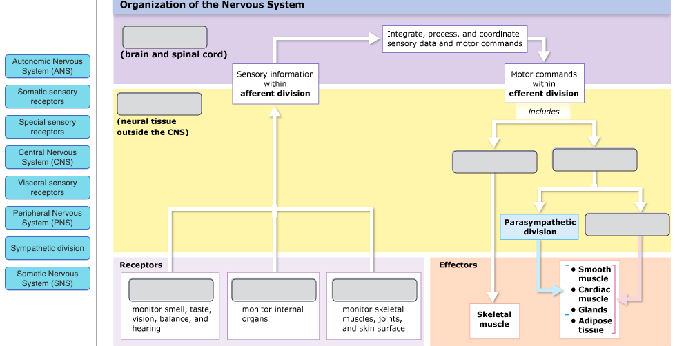
8.1 The Concept of Homeostasis. Homeostasis refers to the body's ability to physiologically regulate its inner environment to ensure its stability in response to fluctuations in external or internal conditions.The liver, the pancreas, the kidneys, and the brain (hypothalamus, the autonomic nervous system and the endocrine system) help maintain homeostasis. Label the divisions and receptors of the nervous system. Part A Drag the labels onto the diagram to identify the divisions and receptors of the nervous system. ANSWER: Correct Art-labeling Activity: The Anatomy of a Multipolar Neuron Identify the structural components of a multipolar neuron. ANSWER:Involuntary division of the afferent nervous system Visceral division of the afferent nervous system Autonomic division of the efferent nervous system. Correct Artlabeling Activity Figure 10.8 Label the anatomic pathway for somatic sensations. Part A Drag the labels onto the diagram ... Drag the labels onto the diagram to identify the peripheral nerves and nerve plexuses: lumbar and sacral plexuses. look at pic What type of neural circuit is indicated by the arrow?
Drag the labels onto the diagram to identify the components of the somatic nervous system. Which anatomical description is TRUE of the sympathetic division of the autonomic nervous system? It is also known as the craniosacral division because of its origins of innervation. Look no further than these interactive, exam-style anatomy quizzes. Learn anatomy faster and. remember everything you learn. Start Now. <. General Structure of a Neuron (Nerve Cell) >. Nose and Nasal Cavity: Openings and Support Structures. Transcribed image text: Prag the labels onto the diagram to identify the components of the autonomic nervous system! Reset Help Cardiac muscle Smooth muscle Brain Ganglionic neurons Preganglionic neuron Visceral Effectors Adipocytes Autonomic nuclei in spinal cord Autonomic nuclei in brain stem Spinal cord Autonomic ganglia Visceral motor nuclei in hypothalamus Glands Preganglionic neuron ... The brain and the spinal cord are the central nervous system, and they represent the main organs of the nervous system. The spinal cord is a single structure, whereas the adult brain is described in terms of four major regions: the cerebrum, the diencephalon, the brain stem, and the cerebellum. A person's conscious experiences are based on ...
Nervous system (anterior view) In this article, we'll discuss the peripheral nervous system and its divisions, as well as the peripheral nerves.The peripheral nervous system (PNS) consists of all the nerves branching out of the brain and spinal cord (the central nervous system, CNS).If you imagine the CNS as the main highway, then the PNS forms all the connecting secondary roads.
Main Difference - Somatic vs Autonomic Nervous System. Peripheral nervous system is the nervous system which is outside the brain and spinal cord. The main function of the PNS is to relay information between the central nervous system and the effector organs. Somatic and autonomic nervous system are the two parts of the peripheral nervous system (PNS).
Print A&P Chapter 11 Fundamentals of the Nervous System and Nervous Tissue flashcards and study them anytime, anywhere.
The nervous system can be divided into two functional parts: the somatic nervous system and the autonomic nervous system. The major differences between the two systems are evident in the responses that each produces. The somatic nervous system causes contraction of skeletal muscles.
Neurotransmitters are an important part of the nervous system. Learn more about the anatomy of the nervous system with our beginner-friendly quizzes and labeled digrams. After crossing the synaptic cleft, neurotransmitters bind to their receptors on the postsynaptic membrane. Once the neurotransmitter binds to its receptor, the ligand-gated ...
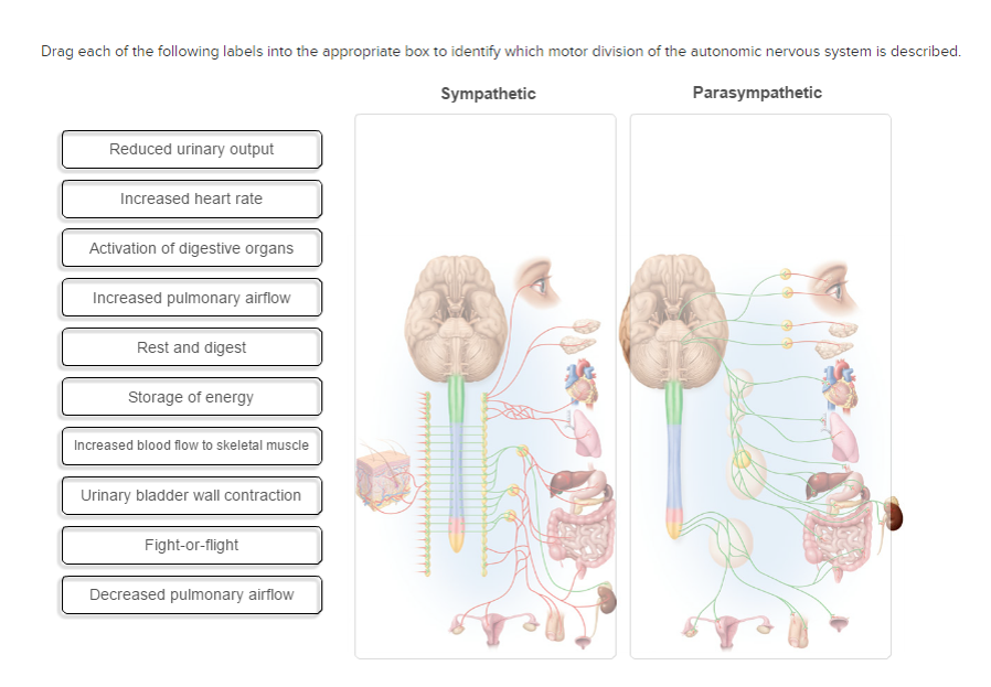
Drag each of the following labels into the appropriate box to identify which motor division of the autonomic nervous system is described. Sympathetic - parasympathetic -
Drag the labels onto the diagram to identify the divisions and receptors of the nervous system. Drag the labels to identify the structural components of a typical neuron. Nice work! You just studied 14 terms! Now up your study game with Learn mode.
Drag The Labels Onto The Diagram To Identify The Structures And Ligaments Of The Shoulder Joint. - Drag The Labels Onto The Diagram To Identify The ... : However, the more mobile a joint, the weaker the joint.. Translation of oppenheim s 1911 paper on dystonia klein 2013. Label the major features of the respiratory system and solved.

Lab Activity Chapter 21 Pdf Lab Activity Chapter 21 Lab Activity Chapter 21 Due 11 59pm On Saturday You Will Receive No Credit For Items You Complete Course Hero
Prag the labels onto the diagram to identify the components of the autonomic nervous system. Drag the labels onto the diagram to identify the components of the integumentary system. Broadly speaking the integumentary system is composed of skin and its appendages subcutaneous tissue deep fascia mucocutaneous junctions and breasts.
Drag the labels onto the diagram to identify the components of the somatic nervous system. What part of the autonomic nervous system is represented in the image? sympathetic division. Which of the following effectors is innervated by neurons that synapse in the collateral ganglia? intestine.
1 1000 2743 Http Uilis Unsyiah Ac Id Oer Files Original Aae4ec936c1d9506d31b432cc5e6891f Jpg Bb77ee2cf8df5299e7c2c0fd03dbc1a1 Dublin Core The Dublin Core Metadata Element Set Is Common To All Omeka Records Including Items Files And
Drag the labels onto the diagram to identify the components of the autonomic ... What part of the autonomic nervous system is represented in the image?
Drag the labels onto the diagram to identify the stages in which the lagging strand is synthesized. Drag the labels onto the diagram to identify the various muscle structures. ... Solved Label The Components Of A Knee Jerk Reflex Part A ... 16 1 Divisions Of The Autonomic Nervous System Anatomy Amp Physiology ...
That is: NFP = GBHP - [CHP + BCOP] = 10 mm Hg. Or: NFP = 55 - [15 + 30] = 10 mm Hg ( Figure 25.4.1 ). Figure 25.4.1 - Net Filtration Pressure: The NFP is the sum of osmotic and hydrostatic pressures. A proper concentration of solutes in the blood is important in maintaining osmotic pressure both in the glomerulus and systemically.

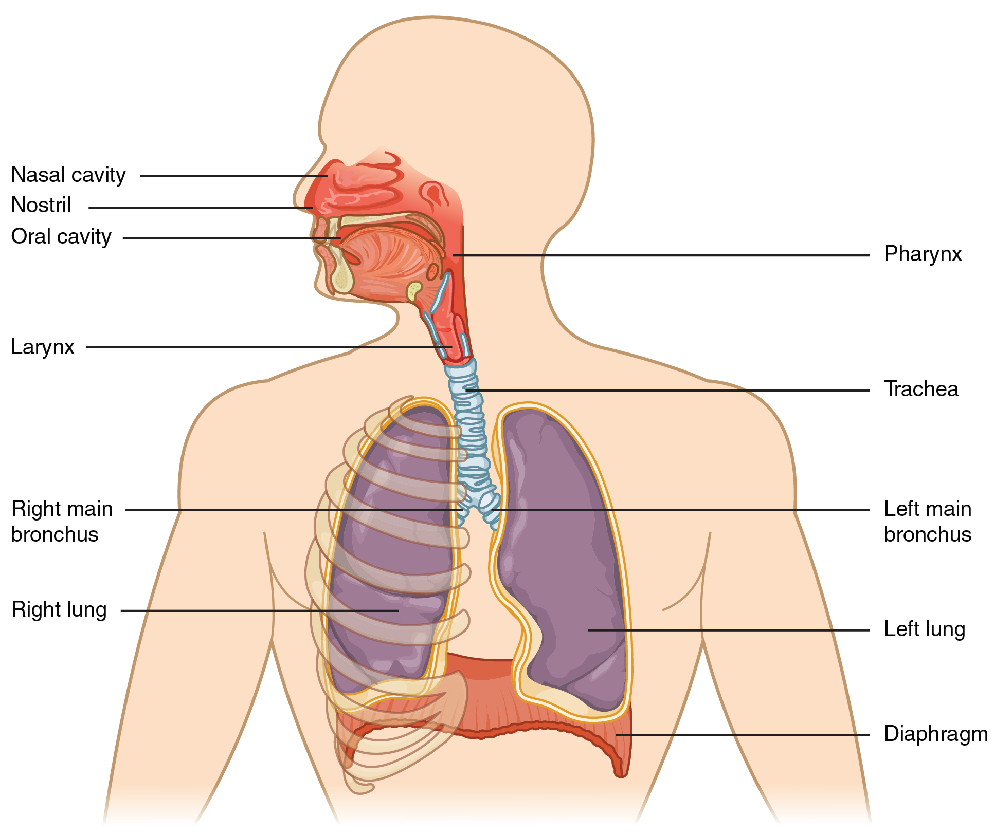
Ou Human Physiology Organs And Structures Of The Respiratory System Ou Human Physiology Textbook Openstax Cnx
Drag the labels onto the diagram to identify the components of the somatic nervous system. Part A Drag the labels onto the diagram to identify the components of the somatic nervous system. Reset Help Brain Somatic motor nuclei of brain stem Somatic motor nuclei of spinal cord Spinal cord Skeletal muscle Upper motor neurons in primary motor ...
Which division of the ANS is referred to as the "rest and digest" ... Click and drag each label on the left to identify the anatomical components of the ...
Drag the labels onto the diagram to identify the origins o the cranial ... onto the diagram to identify the components of the autonomic nervous system.

Drag The Labels Onto The Diagram Of The Axon To Identify Events Occurring During An Action Homeworklib
Art-labeling Activity: The Anatomy of the Ear (External and middle ear) Drag the labels onto the diagram to identify the anatomical structures of the external and middle ear. Reset Help External Ear Middle Ear Internal Ear Tympanic membrane Round window Auricie Tympanie caviy Extem acouste metus Oval window Auditory ossicles Elastic canlage ; Question: Art-labeling Activity: The Anatomy of the ...
Drag the labels onto the diagram to identify the components of the autonomic nervous system. visceral effectors: ( top down) 1. smooth muscle 2. glands. 3. cardiac muscle 4. Adipocytes top middle to bottom middle 1. visceral motor nuclei in hypothalamus 2. autonomic ganglia 3. ganglionic neurons ...

Drag The Labels Onto The Diagram To Identify The Parts Of The Corticospinal Pathway Part A Homeworklib
Part A Drag the labels onto the diagram to identify the sympathetic trunk and the branches of a spinal nerve. ANSWER: Help Reset Spinal cord Dorsal root ganglion Ventral root Sympathetic trunk ganglion Sympathetic trunk Ventral ramus of spinal nerve Gray ramus communicans White ramus communicans ...
• To identify the components of the intrinsic conduction system. • To recognize that the intrinsic conduction system coordinates heart activity by determining the direction and speed of heart depolarization. • To relate heart electrical activity to an ECG wave tracing.
Drag the labels to the correct locations on the. Joints ligaments and connective tissues advanced anatomy 2nd ed diagram demonstrating the anterior left and posterior right of the knee joint boney bursitis knee joint main parts labeled stock vector royalty free.
11 Introduction To The Nervous System And Nervous Tissue Reset help vesicle neurotransmitter calcium channel synaptic terminal synaptic cleft receptor for neurotransmitter correct activity. Drag the labels onto the diagram to identify the various synapse structures. Part a structure of a chemical synapse drag the labels onto the diagram to ...
15.3 Hearing Audition (Hearing) Hearing, or audition, is the transduction of sound waves into a neural signal that is made possible by the structures of the ear (Figure 15.3.1).The large, fleshy structure on the lateral aspect of the head is known as the auricle.Some sources will also refer to this structure as the pinna, though that term is more appropriate for a structure that can be moved ...
Part A Drag the labels onto the diagram to identify the parts of the dissected sheep brain, median section (part 2 of 2). ANSWER: Correct Art-labeling Activity: Figure 17.4a (1 of 3) Part A Drag the appropriate labels to their respective targets.
Drag the labels onto the diagram to identify the divisions and receptors of the nervous system. look at pic Drag the labels to identify the structural components of a typical neuron.
Anatomy of the nervous system. No ligaments connect the bones at this joint. ANSWER Part A Drag the labels onto the diagram to identify the parts and from www.coursehero.com * fibrous structure around the glenoid fossa. • identify the components of a synovial joint. An er diagram for a college system is an entity relationship diagram that is used to identify the entities of the college ...
Division of Human Nervous System (with diagram) Human nervous system is mainly divided into 3 divisions, which are as follows: 1. Central Nervous System 2. Peripheral Nervous System 3. Autonomic Nervous System. The nervous system is mainly divided into central nervous system, peripheral nervous system and autonomic nervous system.
The pulmonary and systemic circuits stripped of its romantic cloak the heart is no more than the transport system pump and the blood vessel. Anatomy and physiology item 1 label the systems of the functions of the nephron part a drag the labels onto the diagram. You can click to make it bigger!
Part a drag the labels onto the diagram of neurochemical communication at an autonomic synapse. Cns central nervous system 7. Drag the labels onto the diagram to identify parts of the neuromuscular junction. What part of the nervous system performs information processing and integration. Drag the labels onto the diagram to identify the components of somatic sensory pathways. By antlab plays quiz not verified by sporcle. Show transcribed image text drag the labels onto the diagram ...
The nervous system has two major parts: the central nervous system (CNS) and the peripheral nervous system (PNS). The central system is the primary command center for the body, and is comprised of ...
The Autonomic Nervous System and Visceral Sensory Neurons15 ... Figure 15.1 Place of the autonomic nervous system (ANS) and visceral sensory components in the structural organization of the nervous system. Central nervous system (CNS) Peripheral nervous system (PNS) ... is mapped to the cervical area in this diagram, all nerves to the periphery ...
Figure 10.2.2 - Muscle Fiber: A skeletal muscle fiber is surrounded by a plasma membrane called the sarcolemma, which contains sarcoplasm, the cytoplasm of muscle cells. A muscle fiber is composed of many myofibrils, which contain sarcomeres with light and dark regions that give the cell its striated appearance.
Drag the labels onto the diagram to identify parts of the neuromuscular junction. Part a drag the labels onto the diagram of neurochemical communication at an autonomic synapse. Drag and drop the descriptive labels of events into the correct sequence at the chemical synapse.

Drag The Labels Onto The Diagram To Identify The Components Of The Autonomic Nervous System Prag Homeworklib
What part of the nervous system performs information processing and integration. Cns central nervous system 7. Anatomy Exam 2 Flashcards Easy Notecards Drag the labels onto the diagram to identify parts of the neuromuscular junction. Drag the labels onto the diagram to identify parts of the neuromuscular junction. First 2 from top to bottom ...








0 Response to "38 drag the labels onto the diagram to identify the components of the autonomic nervous system."
Post a Comment