36 simple squamous epithelium labeled diagram
Microscope Simple Squamous Epithelium Labeled Diagram Written By MacPride Friday, December 25, 2020 Add Comment Edit. Epithelium Web Lab. What Are The Differences Of Simple And Stratified Tissue Sciencing. Epithelial Tissue Anatomy Physiology. Https Www Augusta Edu Scimath Biology Docs Animaltissues Pdf. 4 2 Epithelial Tissue Anatomy And Physiology. Simple Cuboidal Epithelium Tissue Body Tissues Basement. Simple Columnar Epithelium Basement Membrane. Stratified Cuboidal Epithelium Definition And Function Biology. Examining Epithelial Tissue Under The Microscope Human Anatomy. Lab Exercise 4 Epithelial Tissues Connective Tissue Proper What.
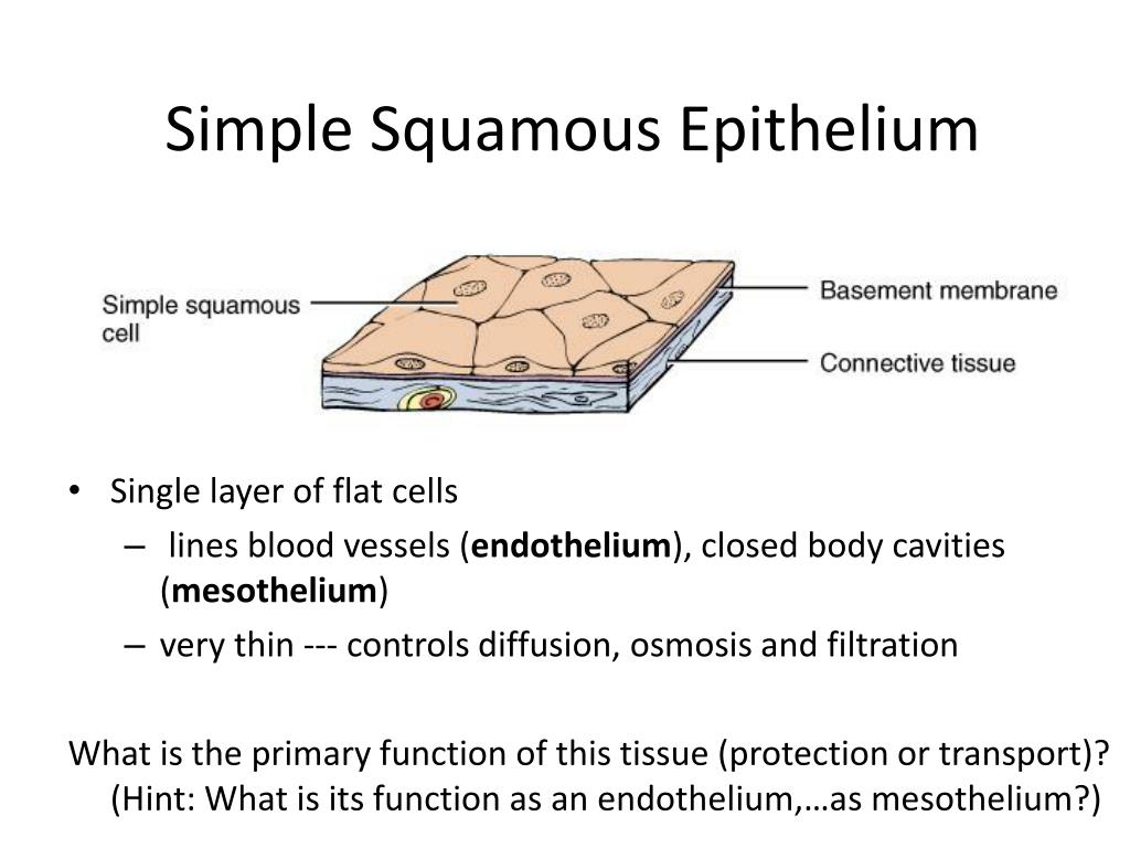
Squamous. Squamous means scale-like. simple squamous epithelium is a single layer of flat scale-shaped cells. Both the endothelial lining of blood vessels and the mesothelial lining of the body cavities are simple squamous epithelium.. Try to identify the simple squamous epithelia in these pictures.
Simple squamous epithelium labeled diagram
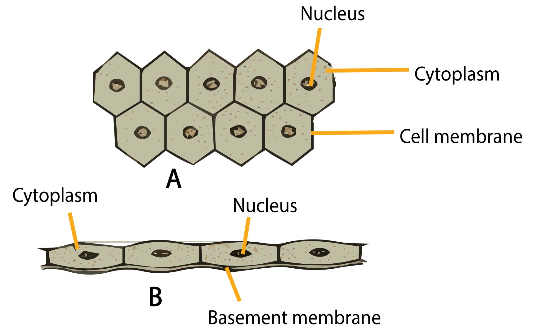
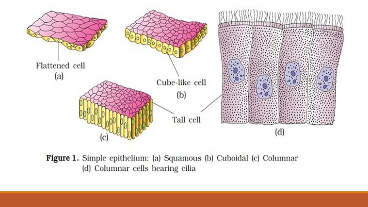
Simple Epithelia Simple Squamous Epithelium (Figure 4.3a) A simple squamous epithelium is a single layer of flat cells. When viewed from above, the closely fitting cells resemble a tiled floor. When viewed in lateral section, they resemble fried eggs seen from the side. Thin and often permeable, this type Simple squamous epithelium is the tissue that creates from one layer of squamous cells which line surfaces. The squamous cells are thin, large, and flat, and consisting of around nucleus. These tissues have polarity like other epithelial cells and consist of a distinct apical surface with special membrane proteins. A. Simple columnar epithelium. Slide 29 (small intestine) View Virtual Slide Slide 176 40x (colon, H&E) View Virtual Slide Remember that epithelia line or cover surfaces. In slide 29 and slide 176, this type of epithelium lines the luminal (mucosal) surface of the small and large intestines, respectively. Refer to the diagram at the end of this chapter for the tissue orientation and consult ...
Simple squamous epithelium labeled diagram. A simple squamous epithelium, also known as pavement epithelium, and tessellated epithelium is a single layer of flattened, polygonal cells in contact with ... The outer wall is composed of a single layer of flat cells (a simple squamous epithelium). Two of the nuclei (stained purple) are indicated by arrows labeled n. Two areas of cytoplasm (stained pink) are indicated by arrows labeled c. The simple squamous epithelium shown here is the outer wall of the glomerular capsule. More information about ... Simple cuboidal epithelium in Mallory stain (longitudinal cut). Note the dark chromatin clumps in the nuclei. Underneath the epithelium lies a small blood vessel filled with orange-colored blood cells. Slide 6 Cross-section of tubules. The smaller ones clustered in the center and upper left are lined by simple squamous epithelium. Simple Squamous Epithelium. Function: Passage of materials by diffusion and filtration. Location: Air sacs of lungs. Simple Cuboidal Epithelium.7 pages
Epithelium is a tissue that lines the internal surface of the body, as well as the internal organs. Simple epithelium is one of the types of epithelium that is divided into simple columnar epithelium, simple squamous epithelium, and simple cuboidal epithelium. Bodytomy provides a labeled diagram to help you understand the structure and function of simple columnar epithelium. Simple squamous epithelium. the cells are flattened and single-layered · Simple cuboidal epithelium · Simple columnar epithelium · Pseudostratified columnar ... Simple squamous epithelium, isolated (400x) Buccal mucosal In the center of this image are two simple squamous epithelial cells that are still attached to each other. Notice that the location of the nucleus (nuc) is in the center of the cell. It is surrounded by the much paler cytoplasm (cyt). The simple squamous epithelium location specifically exists in the lining of the blood vessels like the arteries, veins, and capillaries. It is also found lining the alveoli or air sacs within the ...
Simple squamous epithelium definition. Simple squamous epithelium is a type of simple epithelium that is formed by a single layer of cells on a basement membrane. It is a type of epithelium formed by a single layer of squamous or flat cells present on a thin extracellular layer, called the basement membrane. Epithelial Tissue Diagram. The epithelium is a complex of specialized cellular organizations arranged into sheets without significant intercellular substance. It is a thin, continuous, protective layer of cells. The epithelial tissues perform many functions including protection from abrasion, radiation damage, chemical stress and. Think ... Thyroid gland, 40X objective 400X total magnification, simple cuboidal. Key: N=mucleus. CT= connective tissue. L=lumen It is made up of simple squamous epithelium and a connective tissue layer underneath (lamina propria serosae). A characteristic feature of the ileum is the Peyer's patches lying in the mucosa. It is an important part of the GALT (gut-associated lymphoid tissue). One patch is around 2 to 5 centimeters long and consists of about 300 aggregated ...
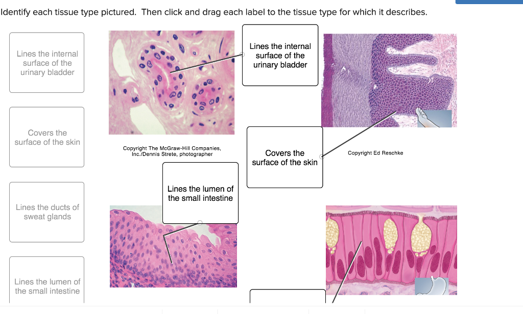
The epithelial lining at the entrance (vestibule) to the nasal cavity exhibits a gradual change from keratinized stratified squamous epithelium of the skin in the nasal vestibule (shown in slide 124F) View Image, to the pseudostratified columnar ciliated epithelium that is characteristic of the nasal mucosa posterior to the vestibule (shown in ...
Form the Outer Covering of the skin and some internal organs. Form the Inner Lining of blood vessels, ducts and body cavities, and the interior of the respiratory, digestive, urinary and reproductive systems. Glandular epithelia. Constitute the secretory portion of glands. Simple squamous epithelium. Most delicate epithelium.
Squamous means scale-like. simple squamous. Bodytomy provides a labeled diagram to help you understand the structure and Simple Columnar Epithelium: Labeled Diagram and Function. Epithelium is a tissue that lines the internal surface of the body, as well as the internal organs. Simple epithelium is one of the types of epithelium that is.
Simple squamous epithelia consist of a single layer of flattened cells. This type of epithelia lines the inner surface of all blood vessels (endothelium), forms ...
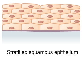
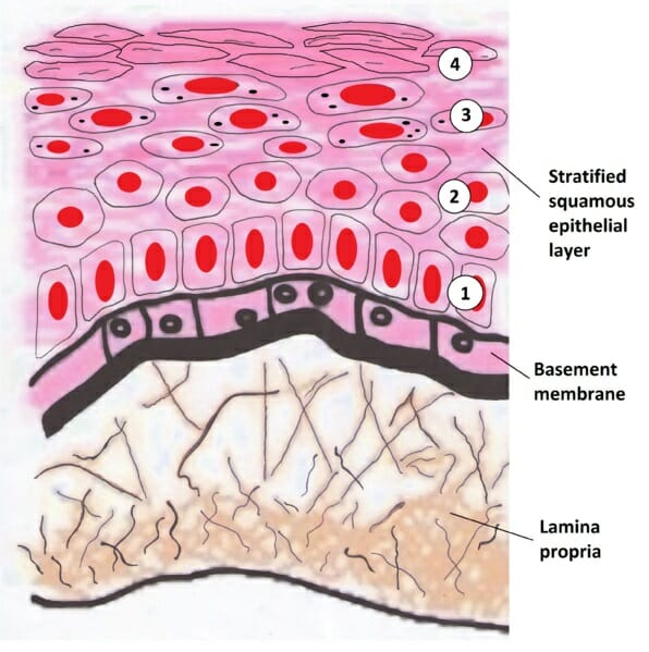
A stratified squamous epithelium is a tissue formed from multiple layers of cells resting on a basement membrane, with the superficial layer(s) consisting of squamous cells. Underlying cell layers can be made of cuboidal or columnar cells as well.
Simple Squamous Epithelium (Diagram) Stratified Squamous Epithelium (Diagram) Simple Cuboidal Epithelium (Diagram) Stratified Cuboidal Epithelium (Diagram) ... Cranial Nerves-Anatomy 12 Terms. kajcolarusso TEACHER. Peripheral Nerves 18 Terms. kajcolarusso TEACHER. THIS SET IS OFTEN IN FOLDERS WITH... Epithelial Basics 10 Terms.
by A Rani · 2014 — EPITHELIAL TISSUE or EPITHELIUM. • The basic tissue of the body. ... Simple Squamous Epithelium. • Single layered. • Flat cells.32 pages
Stratified, keratinized squamous epithelium. Image: Epidermis-structure diagram labeled. By BruceBlaus, License: CC BY 3.0. The outermost cell layers of the epithelium consist of flattened cells with no nuclei and cytoplasm, converting into scales. They are called the stratum corneum, and their
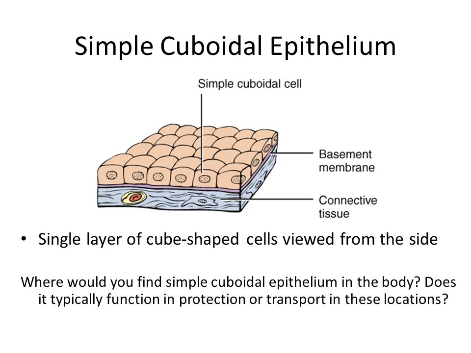
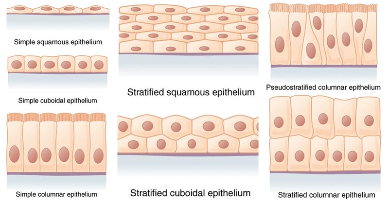
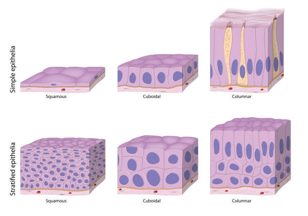
Types of Epithelial Tissue. There are three types of epithelial cells, which differ in their shape and function. Squamous- thin and flat cells Cuboidal- short cylindrical cells, which appear hexagonal in cross-section Columnar- long or column-like cylindrical cells, which have nucleus present at the base On the basis of the number of layers present, epithelial tissue is divided into the simple ...
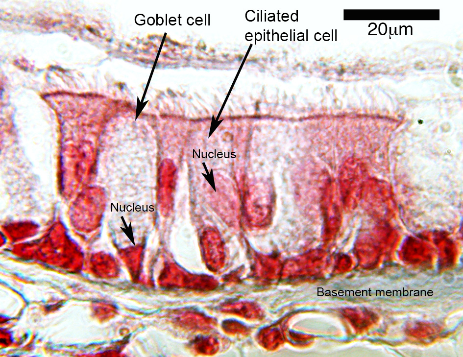
Simple Columnar Epithelium Labeled Diagram. Ciliated columnar epithelium is composed of simple columnar epithelial cells with cilia on their apical This illustration shows a diagram of a goblet cell. These labelled diagrams should closely follow the current Science (simple squamous epithelium).
Simple squamous epithelium, c.s. (400X) thin section Kidney cortex The arrows in the image point to the nuclei of simple squamous epithelial cells. This image was made from a thin section of the kidney at the same magnification as the previous image (400X). It is about one-fifth to one-tenth the thickness of the slides used to make the top ...
Sep 2, 2021 — Figure 1 shows a diagram of simple squamous epithelium labeled. The tissue is polarized with one surface that faces the external environment ...
Simple epithelium can be divided into 4 major classes, depending on the shapes of constituent cells. The cells found in this epithelium type are flat and thin, making simple squamous epithelium ideal for lining areas where passive diffusion of gases occur.Areas where it can be found include: skin, capillary walls, glomeruli, pericardial lining, pleural lining, peritoneal cavity lining, and ...
These labelled diagrams should closely follow the current Science courses in histology, anatomy and ... squamous epithelium) ORIGIN: ectoderm lamina propria ... Covers the external surface. blood vessel lined by endothelium (simple squamous epithelium) ORIGIN: mesoderm lumen GENERALISED SECTION epithelium OF THE BODY connective tissue beneath ...
Simple Squamous Epithelium Definition. Simple squamous epithelia are tissues formed from one layer of squamous cells that line surfaces. Squamous cells are large, thin, and flat and contain a rounded nucleus. Like other epithelial cells, they have polarity and contain a distinct apical surface with specialized membrane proteins.
FIGURE 1-1 (a) Simple squamous epithelium lines the lumina of vessels, where it permits diffusion.(b) A photomicrograph of this tissue and (c) a labeled diagram.Simple squamous epithelia that line the lumina of vessels are referred to as endothelia, and that which cover visceral organs are referred to as mesothelia.
Stratified squamous epithelium is the most common type of stratified epithelium in the human body. The apical cells appear squamous, whereas the basal layer contains either columnar or cuboidal cells. The top layer may be covered with dead cells containing keratin. The skin is an example of a keratinized, stratified squamous epithelium.
A. Simple columnar epithelium. Slide 29 (small intestine) View Virtual Slide Slide 176 40x (colon, H&E) View Virtual Slide Remember that epithelia line or cover surfaces. In slide 29 and slide 176, this type of epithelium lines the luminal (mucosal) surface of the small and large intestines, respectively. Refer to the diagram at the end of this chapter for the tissue orientation and consult ...
Simple squamous epithelium is the tissue that creates from one layer of squamous cells which line surfaces. The squamous cells are thin, large, and flat, and consisting of around nucleus. These tissues have polarity like other epithelial cells and consist of a distinct apical surface with special membrane proteins.
Simple Epithelia Simple Squamous Epithelium (Figure 4.3a) A simple squamous epithelium is a single layer of flat cells. When viewed from above, the closely fitting cells resemble a tiled floor. When viewed in lateral section, they resemble fried eggs seen from the side. Thin and often permeable, this type














0 Response to "36 simple squamous epithelium labeled diagram"
Post a Comment